Listen to this story:
0:00
/
For decades, scientists have been teased by the strange but inaccessible cephalopod visual system. Now, thanks to a technological breakthrough from a lab in Oregon, data are finally coming straight from the octopus brain.
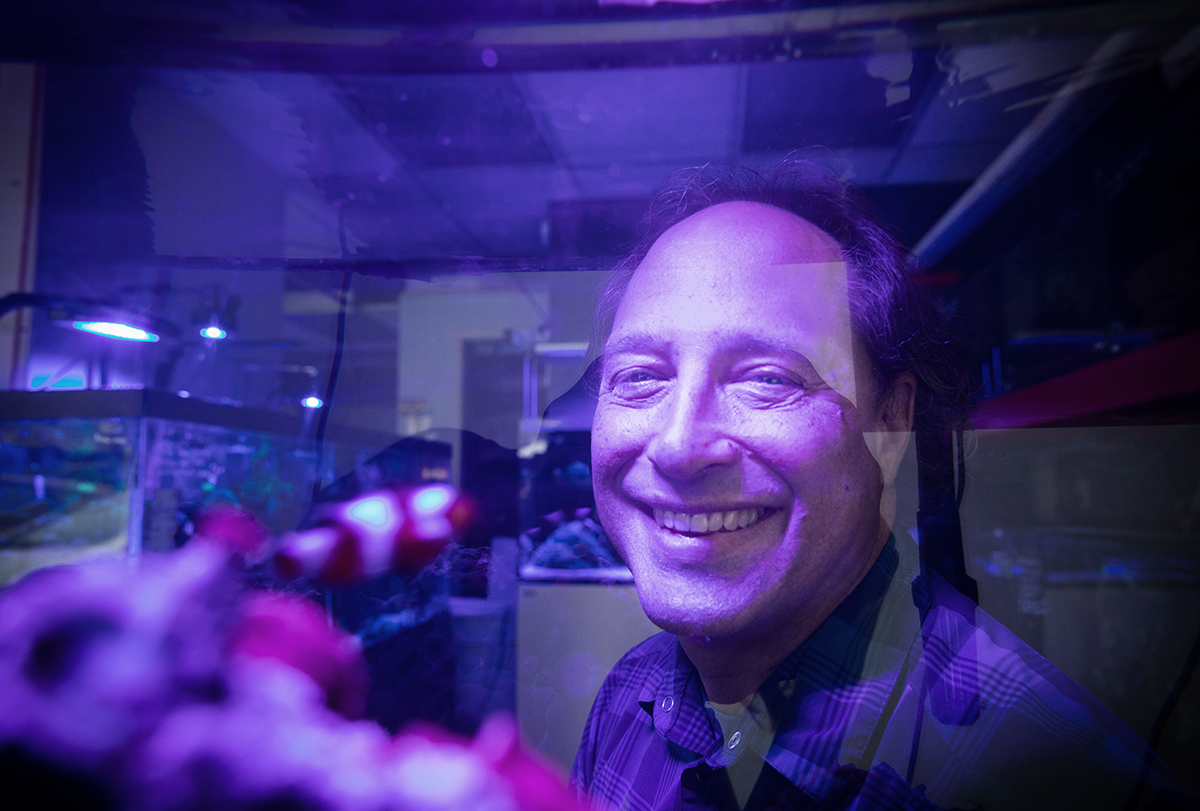
The Niell Lab’s research interests are apparent the moment you enter the team’s space. An eyeball balloon hangs from the ceiling, and a relief sculpture of a bloodshot, veiny eye peers into the lab from above the door. Then there are the octopuses: plush toy ones and figurines and wall prints, as well as 3D-printed versions of their brains scattered around the place.
Cristopher Niell, professor of biology at the University of Oregon, has spent his career studying vision in different species and settings, and he is “a little bit allergic” to doing what everyone else is doing, he says. For instance, rather than sticking to the field’s classic animal models (cats and nonhuman primates), he helped develop the zebrafish and mouse as new models.
But he came to the octopus thanks to Judit Pungor, now a staff scientist in his lab. She has loved the animal and other cephalopods since she was 5 years old, visiting the Monterey Bay Aquarium in California with her parents. There she saw a cuttlefish, a master camouflager, vanish into the background, and “it made my heart stop,” she says.
Since an impromptu collaboration in 2010, the pair has been trying to crack the octopus visual system. Only three groups in the animal kingdom use their vision to detect objects on a regular basis: cephalopods, arthropods and vertebrates. Vision in the latter two is well studied, says Dan-Eric Nilsson, professor emeritus of sensory biology at Lund University. Scientists recorded from the visual cortex of mammals and figured out how photoreceptors in the eyes respond to light and how brain regions in the circuit extract details from the scene.
But every attempt to record from neurons in cephalopods’ optic lobe—and thus understand how the system truly works—failed because of technical hurdles.
“So there is a big blank spot there that really needs to be investigated,” Nilsson says.
The gap is not due to lack of interest. Biologists in the 20th century discovered that octopuses have eyes like those of vertebrates and brains larger than any other invertebrate, yet with the neural hardware of an invertebrate. They also cataloged how octopuses use their excellent vision to direct behavior.
But that’s where the road has ended. Octopuses are separated from humans by more than 500 million years of evolution. Their last common ancestor did not have vision, and so any similarities between the two are likely driven by a key function rather than “family resemblance,” says Paul Katz, professor of biology at the University of Massachusetts Amherst. Katz thinks studying cephalopods is “seriously the most important thing that neuroscience could do right now,” and studying cephalopod vision in particular, he adds, could reveal the forces that shaped the sense.
Niell, Pungor and their team have finally figured out how to do it.
A
s a Ph.D. student at Stanford University studying the anatomy of the octopus visual system, Pungor, like the cephalopod researchers before her, wasn’t satisfied with only anatomical or behavioral information. She wanted to know what the neurons were doing.The layout of the octopus visual system made this difficult. In vertebrates, photoreceptors in the back of the eye respond to visual stimuli; several layers of other cell types in the retina process that signal before it exits the eye and travels to the thalamus and cortex for further processing. In octopuses, however, the signal from the photoreceptors immediately exits the eye, so initial processing takes place in the outer layers of the optic lobe.
Pungor managed to record from cells in the back of the eye, which scientists in the 1960s and ’70s had also done. But recordings in the optic lobe never worked. The region, like the rest of the octopus brain, contains many tiny cells packed together, Pungor says, which made it “insurmountable” to isolate the signal of a single neuron from all the background noise.
One of Pungor’s advisers recommended she reach out to Niell, then a postdoc at the University of California, San Francisco. He had recorded from the mouse visual cortex using multisite silicon probes, which might work better than her attempts to record with one electrode at a time, her adviser reasoned. She sent Niell a email asking to collaborate, and Niell, after skimming the octopus literature, realized the animal was a “completely unsolved area of vision,” he says.
That discovery was like a siren call to him. The vision field had formed several core principles from work in mammals such as cats, mice and nonhuman primates, and some of these had held up in insects. David Hubel and Torsten Wiesel demonstrated in their Nobel-Prize-winning experiments that neurons in the primary visual cortex are tuned to edges in the visual scene, such as boundaries between dark and light, rather than to individual pixels like in the retina. Later experiments in the field mapped how the brain transforms visual information each step of the way as it builds a scene: first as pixels, then edges, and later as textures, motion, shapes and faces.
“Each stage is basically extracting something different rather than just sending the same image along,” Niell says. He wanted to know if it was the same in the octopus, and if so, how the system managed to perform the same type of computations with such different circuitry.
Pungor drove two hours from Stanford’s marine station near Monterey to San Francisco with a bucket of freshly caught squid buckled into her passenger seat. The pair tried to record from the squid for a week, but all of their experiments failed. The signal was too noisy to parse apart. They went their separate ways, Niell enthralled with the science but needing to devote time to his job search, and Pungor quietly worried that she had wasted his time.
Five years later, though, they caught up over dinner when Niell gave a seminar at the University of Chicago, where Pungor was finishing up a postdoc. During that time, Pungor had helped sequence the first cephalopod genome, and Niell had started his lab at the University of Oregon. Now he was close to tenure, a little bored and ready to take a risk. They agreed it was time to try again.
Pungor flew out to Oregon for another round of experiments and to interview for a position in Niell’s lab. This time, they tried calcium imaging. Typically, these experiments take place in genetically modified animals designed to express a protein that glows when it binds to calcium. Niell had previously used this technique during his Ph.D. while studying vision in zebrafish. The genetic version was not fully developed at the time, so Niell used a method called “bolus-loading”—injecting a large amount of a calcium indicator dye into the brain so it is taken up by cells.
They placed a rice-sized squid in a petri dish and poured calcium indicator dye over it. Because the squid was so small, they hoped the dye could seep into the mantle and infiltrate the nerves inside. The squid was motionless; nothing happened at first. But when it jetted across the dish, the animal lit up “like gangbusters,” Pungor says. The two had shown that calcium imaging can capture neural activity in cephalopods.
That gave them “a tool that can begin to answer these questions,” Pungor says. She accepted the position in Niell’s lab and got to work converting it from a mouse-only to a mouse-and-octopus lab. Unraveling the octopus visual system didn’t seem so impossible anymore, though it would take them seven more years to iron out their protocol.
P
ungor doesn’t remember the exact date that she first captured visual responses from the cells—“it was probably like 2015 or 2016,” she says—but she does remember it was a Wednesday night at 8 pm. She ran through the hallway of the empty building, yelling with joy, on the hunt for someone to celebrate with, until she ran into a graduate student from another lab in his office and shared her good news.But there are many ways that experiments in cephalopods can go wrong—so many that Pungor created a “spreadsheet of doom,” she says, with more than 50 columns to track parameters, such as the starting and ending temperature and pH of the water, the volume of calcium indicator injected and the recipe of different solutions. Often, everything would seem to go smoothly, but the optic lobe neurons would not respond to visual stimuli.
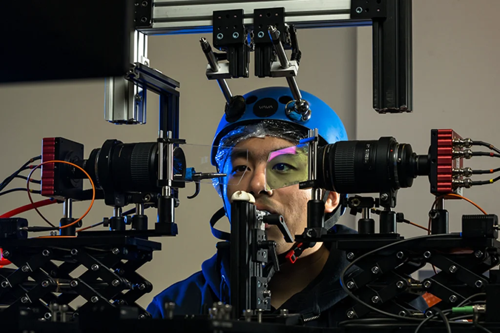
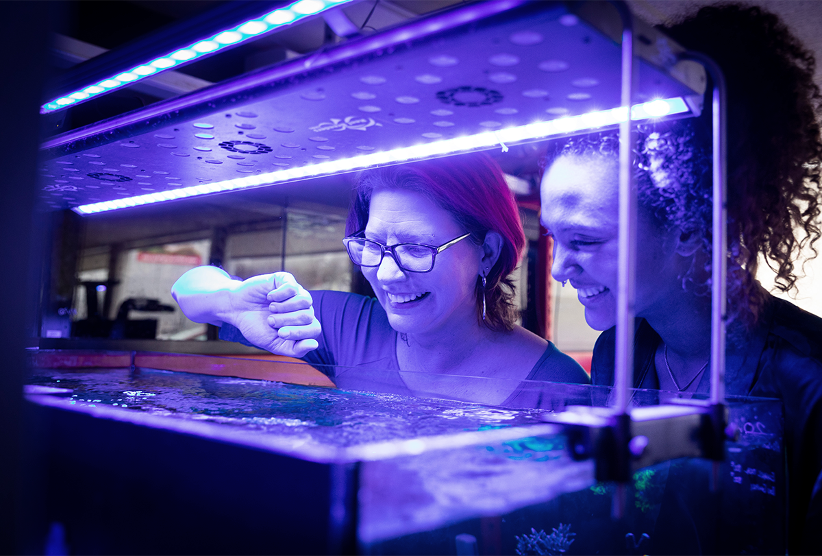
By 2022, Pungor and graduate student Angelique Allen had eked out the right parameters and were consistently recording visual responses in the octopus optic lobe. They study Octopus bimaculoides, also known as the California two-spot octopus, which grows to be about 45 centimeters long. The experiments take place in “the brain box,” a black, 3D-printed apparatus filled with cold saltwater that’s like a miniature movie theater for an octopus eye: A small raised platform inside the main compartment serves as a front-row seat for an isolated eye and brain; a slit across from the platform holds a smartphone-sized LCD screen that plays black-and-white movies of flashing squares, migrating dots and sweeping bars while Allen uses calcium imaging to record activity in the brain tissue. The screen has a removable filter so Allen can measure responses to either brightness or polarization—octopuses are likely color-blind but can detect different angles of light as it bounces off objects.
Once the preparation is ready, Allen injects the calcium indicator into two spots in the optic lobe. She covers it in agarose to keep everything still and seats the coverslip on the platform in the brain box. She holds a mirror between the LCD screen and platform to ensure the preparation is positioned correctly and “makes eye contact with the eye” to check that the pupil is pointed at the screen.
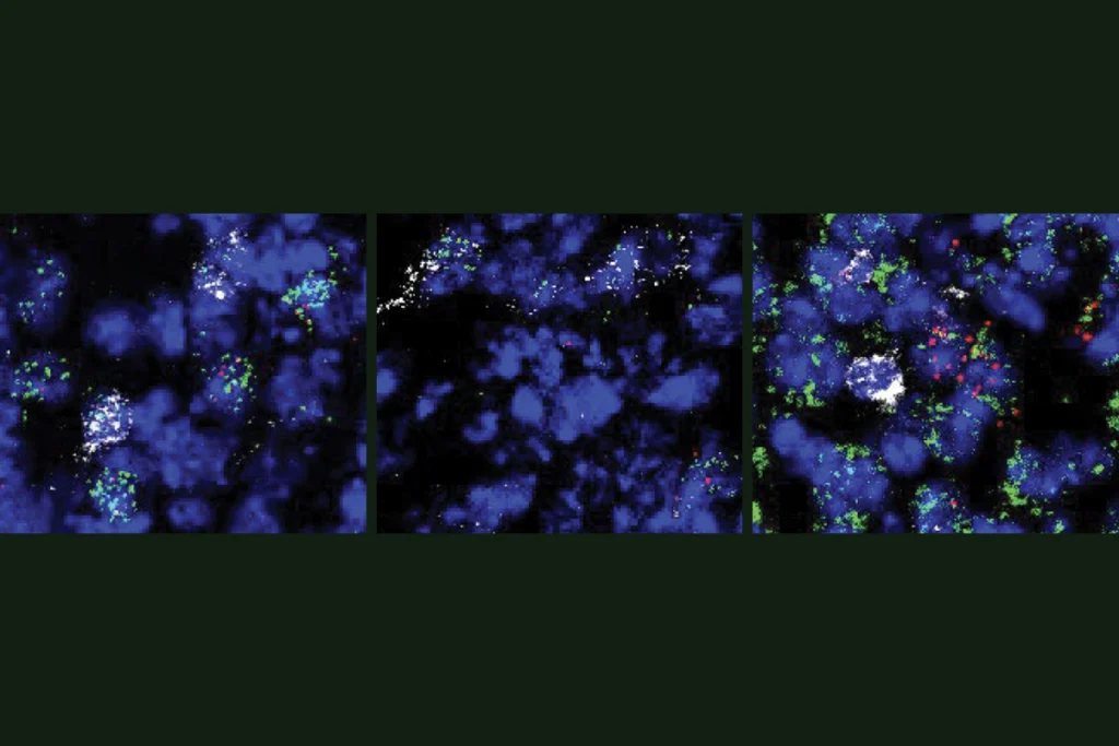
Allen secures the brain box under the two-photon microscope, closes the curtains around the rig, takes a seat at the computer and plays the first movie. Before she starts collecting data, she flashes a pulse of light to check that the cells respond to visual stimuli. On the scope computer screen, the optic lobe glows faint green, peppered with sharper clusters of circular cell bodies. After the flash of light, a wave of brightness creeps across the optic lobe, like the afterglow of fireworks.
Allen then plays the black-and-white movies for the tissue preparation and records the resulting activity. After she processes the images in a data analysis program, the cell’s activity looks more like the twinkling of string lights.
T
hrough these experiments, Niell and his team have sketched out a basic framework for how visual processing takes place in the octopus.The optic lobe has several characteristics in common with vertebrate visual systems, the calcium imaging experiments show. Some neurons in the optic lobe respond to increases in light, a property known as on-responsive, whereas others respond to decreases in light, which is known as off-responsive. In addition, the neurons are organized retinotopically, meaning cells that are tuned to adjacent areas of the visual field sit next to each other in the brain, too.
“A completely different pathway through evolution also found this solution of, ‘Oh, let’s build a map of the world in the brain,’” Niell says. “It really points out that visual systems work best when information is organized spatially.”
The response properties change as you move deeper into the optic lobe: Most of the off-responsive neurons are found in the deeper layers, and the on-responsive neurons in that area show a greater preference for smaller stimuli. This difference in selectivity is evidence that each step of the visual pathway extracts different information from the visual input; the input is transformed rather than passively shuttled along, just like in mammals.
So far, both findings align with how vision works in vertebrates and arthropods, and they provide a “pretty good sense” of how the optic lobe works on a population level, Niell says. Now that the researchers have an outline, they want to fill in the details, such as what the individual neurons are doing, what they are tuned to—perhaps orientation, or the direction of motions, like they are in vertebrates—and how they process the polarization angle of light.
“The cephalopod brain was this black box, and they opened the box,” says Sam Reiter, associate professor at the Okinawa Institute of Science and Technology, who studies computational neuroethology. After the failed attempts in the 20th century, the field sustained “rumors and dogma” that performing physiological experiments on the cephalopod brain was impossible, he says. Work from Niell’s group shattered that perception. “It was much more than just they got calcium imaging to work. They showed that this brain could be studied.” Reiter cites the team’s project as motivation to begin his own in-vivo electrophysiology experiments in squid and other species.
The data Niell and his team have collected so far help to “fill in some of the gaps” in how the octopus visual system performs its computations, says Elke Buschbeck, professor of biological sciences and neuroscience at the University of Cincinnati. Adding this previously unstudied animal to the field’s repertoire is valuable, she adds, “because when you find common principles, that really shows you what the important key components are.”
The team has refined the imaging protocol to the point that the data are clean enough to analyze what each cell is responding to rather than analyzing only group-level activity. So far, they are “seeing interesting types of selectivity,” Niell says. The group also created a cell atlas of the optic lobe with single-cell RNA sequencing, so once they have genetic tools, they can examine the relationship between a cell’s molecular identity and selectivity and explore what happens when different subtypes are turned off.
They have some further-off questions, too, such as how the visual system comes together during development, if octopuses process polarization in the same way that other animals process color, and how octopuses use their polarization vision.
Even now, the team’s experiments don’t always work, and they can’t always figure out why, though it’s easier to stomach setbacks now. Pungor often thinks about how long the field has wanted to record from the octopus optic lobe, she says, and how many people have tried and failed. And now here she is, “little old me, sitting in Eugene, Oregon,” recording from the lobe every day. “It’s really humbling.”