Twenty mouse models of autism can be sorted into two subtypes based on functional connectivity across the entire brain, a new preprint reports. The subtypes reflect changes in different molecular pathways and map on to nearly one-quarter of autistic people represented in a large dataset.
The work “moves the needle,” says Kaustubh Supekar, clinical associate professor of psychiatry and behavioral sciences at Stanford University, who was not involved in the study, and is a “first step” toward understanding autism heterogeneity.
The diversity of autism has made it challenging to study its biology and develop treatments. Many papers have grouped people with autism into different subtypes based on their phenotypes, but “these are just statistics-based approaches,” says study investigator Alessandro Gozzi, senior researcher at the Istituto Italiano di Tecnologia. “There is no biologically grounded validation for this.”
So, Gozzi and his team hunted for subtypes in mouse models—a system in which they could probe biological mechanisms—and then examined whether people could be sorted into similar groups. They used functional MRI (fMRI) to measure the global functional connectivity, or synchrony between active regions, across the entire brain in 17 mouse models with genetic alterations linked to autism. They also assessed a mouse model that involved maternal inflammation, another linked to idiopathic autism, and a third featuring an altered gene associated with microglia function.
The emphasis on functional data sets the project apart from previous neuroimaging studies of autism mouse models that focused on structural data, says Ravi Menon, professor of medical biophysics and medical imaging at the University of Western Ontario, who was not involved in the work.
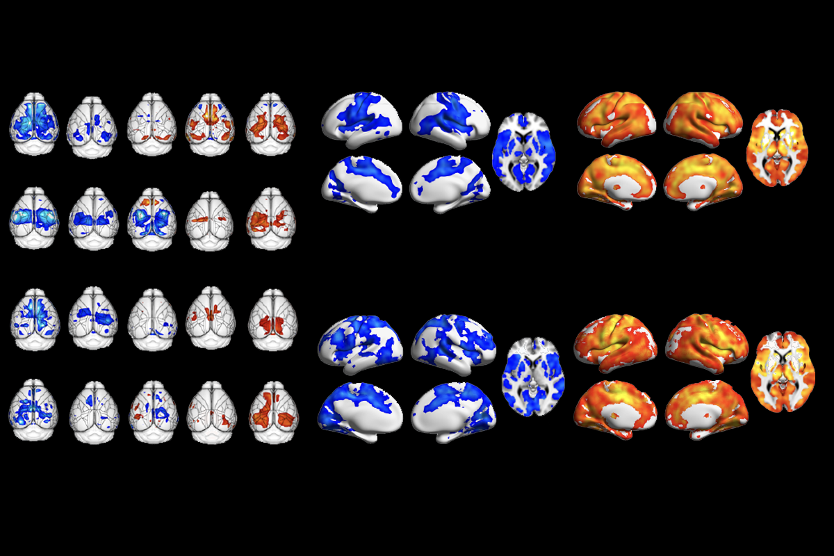
The models broke down into hyperconnectivity and hypoconnectivity subtypes, based on the dominant pattern across brain regions. Active brain regions are over-synchronous in the hyperconnectivity subtype and out-of-sync in the hypoconnectivity subtype, compared with their synchrony in wildtype littermates.
Some brain regions—such as the medial prefrontal cortex, striatum and basal forebrain—were implicated in opposite ways in both subtypes. Others showed subtype-specific changes, including the hippocampus and amygdala in the hyperconnectivity group, and the hypothalamus and somatomotor cortex in the hypoconnectivity group.
T
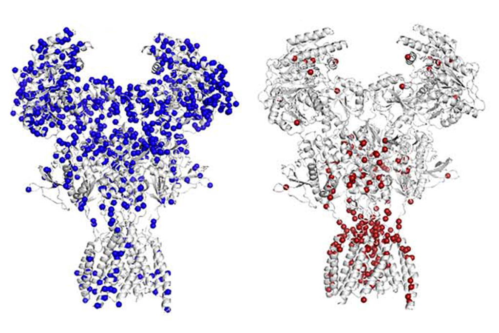
o identify potential biological mechanisms behind each subtype, the researchers built a list of every gene that interacts with the mutated or knocked-out genes for each model, and every protein that interacts with those genes, and removed the ones that the two groups share. “Then you get this mega-group of proteins, and we can see what pathways are there represented beyond chance level,” Gozzi says. The hyperconnectivity group has dysfunctional immune and transcription pathways, while the hypoconnectivity group displays impairments in synaptic function.The mouse-based subtypes can explain some of the variation in people, too. About 24 percent of fMRI scans from 940 people aged 5-30 with idiopathic autism—a majority of which came from the Autism Brain Imaging Data Exchange dataset (ABIDE)—can be sorted into a hyper- or hypoconnectivity subtype, compared with neurotypical controls, which is “quite a strong effect,” Menon says.
That level of cross-species translation came as a surprise, Gozzi says. “As a mouse researcher, what I expected was, like, zero [percent].” And the same types of pathways are altered in people, too, according to an analysis of the two connectivity patterns, using the Allen Institute Human Brain Atlas and postmortem brain tissue from people with autism: synaptic pathways in the hypoconnectivity subtype and immune pathways in the hyperconnectivity subtype.
The comparative aspect of the work “helps us validate and feel confident about our mouse models,” says Janel Le Belle, associate adjunct professor of neurosurgery at the University of California, Los Angeles, who was not involved in the study. “It’s very, very helpful to see this successful translation.”
O
ne limitation of the work is that it does not characterize the subtypes’ clinical profiles, Supekar says. Instead, the researchers compared scores on the Autism Diagnostic Observation Schedule, Second Edition (ADOS-2), a measure of trait severity, and found that people in the hyperconnectivity subtype had more severe traits than people in the hypoconnectivity subtype.Another connectivity-based subtyping study from 2023 clustered people with autism into four subtypes, each with a different clinical profile. For example, one group displayed increased repetitive behaviors and another had greater social challenges. It’s difficult to compare the two groups of subtypes without the same level of clinical detail for the mouse-based work, says Amanda Buch, a postdoctoral researcher at Weill Cornell Medicine in Conor Liston’s lab who worked on the 2023 study.
This type of detailed clinical characterization is not possible with the ABIDE dataset, however, because the phenotypic data were collected in different ways, says Adriana Di Martino, research director for the Autism Center at the Child Mind Institute, who created the dataset and is an investigator on the new study. In the past, critics have flagged these issues as a significant constraint of that dataset. “There’s a need, a desperate need, for more extended, harmonized and deep phenotypic information” to make the search for subtypes “exponentially more fruitful.”
Buch says she also would like to see if different clustering methods pull out the same two subtypes. “Most clustering methods always find clusters,” she says, so finding the same subtypes using different algorithms would strengthen the findings.
It would be interesting to see how the two subtypes change over the course of development, Supekar says. Functional connectivity can, in fact, dramatically change—in some longitudinal studies, children with hyperconnectivity become hypoconnected when they grow older.
“You’re associating a permanent signature, or permanent label, to this individual based on their current connectivity pattern,” which can be flexible, Supekar adds. “But the inside biology is going to be the same.”
Gozzi is interested in this question, too, he says, and is currently working on a project related to developmental changes of connectivity in mouse models. He also would like to use more mouse models—perhaps 80 or 90—to create the “next branch” of subtypes within the broader hypo- and hyperconnectivity buckets.