Autistic people show synaptic differences in brain areas involved in cognitive functions, according to a new study. The work bolsters the hypothesis that an imbalance between inhibitory and excitatory signals in the brain underlies some autism traits.
“This study gives us good confirmation about the excitation/inhibition hypothesis,” says Chirag Mehra, a pediatrician and Ph.D. candidate in Declan Murphy’s lab at King’s College London, who was not involved in the work. “It is helping us understand the neurobiology [of autism].”
Autistic people have more excitatory neurons and a higher density of dendritic spines, which receive signals from other neurons, than non-autistic people do, previous research has found.
Excitatory synapses are typically located on dendritic spines, so higher spine density suggests more excitatory synapses. The new study looked directly at synapse numbers in postmortem brain tissue from autistic and non-autistic people.
“We demonstrated that in [autistic people] there is an alteration in excitation and inhibition, not just because of the number of cells but because of the synapses themselves,” says lead investigator Verónica Martínez-Cerdeño, professor of pathology at the University of California, Davis.
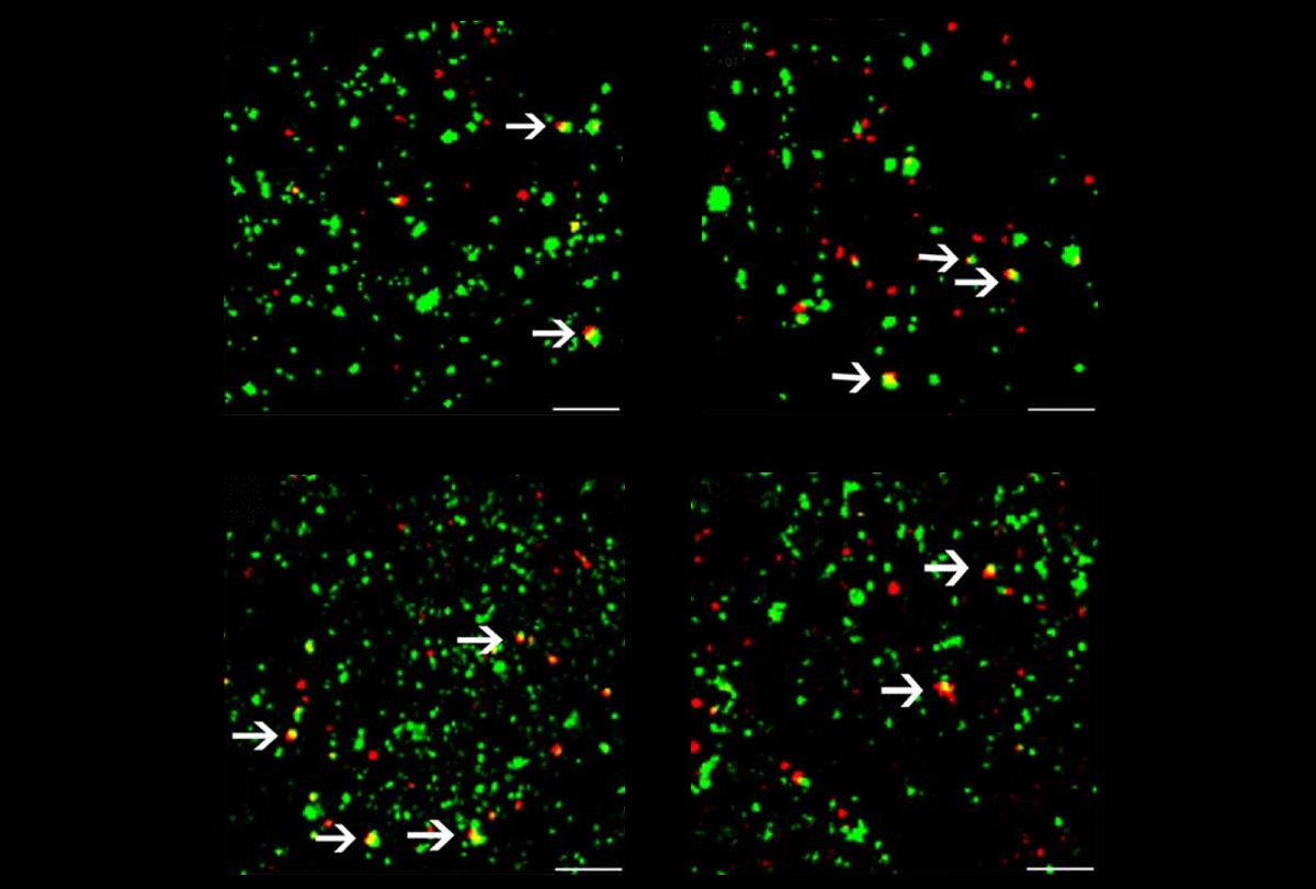
Martínez-Cerdeño and her team analyzed part of the prefrontal cortex, which is involved in processes such as learning and memory, from 10 autistic and 10 neurotypical people, aged 7 to 46 years. The researchers stained the tissue for marker proteins specific to either excitatory or inhibitory synapses.
Compared with non-autistic people, those with autism had more excitatory synapses in the upper layers of the prefrontal cortex and fewer inhibitory synapses across all cortical layers.
T
he increase in excitatory synapses supports earlier research indicating that there are elevated numbers of excitatory cells in the prefrontal cortex of autistic people. The decrease in inhibitory synapses correlates with disruptions in GABA—a neurotransmitter responsible for cortical inhibition—and is consistent with previous findings of fewer inhibitory neurons in mouse models of autism.The imbalance in the number of excitatory and inhibitory synapses in the prefrontal cortex may affect broader brain function and contribute to some of the cognitive and behavioral traits of autism, Martínez-Cerdeño says. The team published their findings in Cerebral Cortex in May.
The work builds on previous research showing dendritic spine alterations in autism, but it goes deeper by differentiating between excitatory and inhibitory synapses, says Alessandro Gozzi, senior researcher at the Istituto Italiano di Tecnologia, who didn’t take part in the study. “It is an interesting confirmation of the fact that synaptic alterations are key to autism,” he says.
Previous work by Gozzi and his team has suggested that the hyperactivity of a protein involved in autism—and the resulting excess of synapses—causes brain connectivity problems in some autistic people. “We showed that when there is an overabundance of synapses, especially excitatory [ones], the brain is hyper-connected,” Gozzi says. The new study, he adds, supports this model, indicating that alterations in neuronal activity may be linked to broader changes in brain connectivity.
But, Mehra says, the study’s focus on a single brain area limits the generalizability of its findings across the entire brain. What’s more, he says, the work does not identify which autism traits are linked to increased excitatory or decreased inhibitory synapses, nor does it clarify whether these measures are causally linked to the condition or if they represent adaptations or compensatory mechanisms in the brain.
M
ehra’s own unpublished research has found that autistic adults have lower synaptic density in brain regions implicated in the condition—a finding that aligns with the fewer inhibitory synapses reported in Martínez-Cerdeño’s work. Mehra and his colleagues used a radioactive tracer that binds to a synaptic vesicle protein and a brain-scanning method called positron emission tomography to gauge synapse number across the entire brain in 12 autistic and 27 non-autistic adults.In some autistic people, a lower synaptic density is associated with difficulties in social communication, according to unpublished results that Mehra presented in May at the 2024 International Society for Autism Research annual meeting in Melbourne, Australia.
“Our study is telling us that synapses are important, and we may possibly see measurable differences in synaptic number,” he says. But that work doesn’t distinguish between excitatory and inhibitory synapses.
Future work on the number of synapses in autism, Mehra adds, may help us to understand brain connectivity differences in people with the condition.