Space cadets
People with autism are better able to visualize objects rotating in space — perhaps because their brains are wired differently than healthy controls.
-
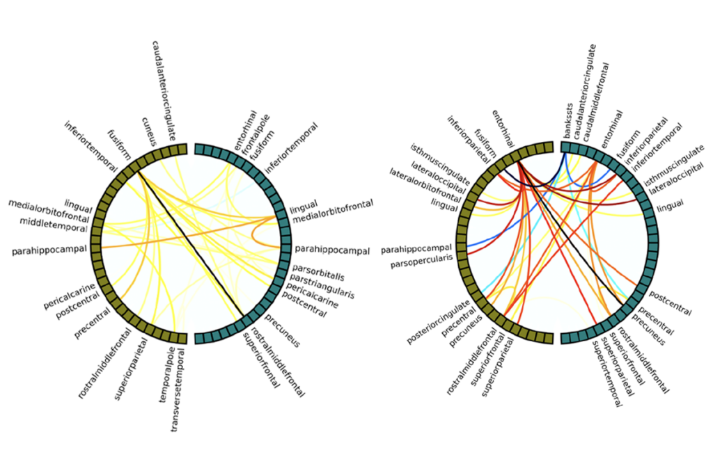
White’s anatomy: Diffusion tensor imaging — which traces the flow of water molecules through the brain — shows abnormalities in white matter in the brains of people with autism compared with controls.
People with autism show a clear advantage in a task that requires them to imagine rotating an object in space, according to results presented Friday in a poster session at the Wiring the Brain meeting held outside Dublin, Ireland.
However, imaging results suggest that this advantage may be the result of structural and functional abnormalities in brain connectivity in those individuals.
In the study, researchers scanned the brains of 22 young adults with high-functioning autism and 22 typical controls matched for age and IQ as they looked at two sets of L-shaped objects on a black background.
In both sets of images, two objects are aligned side by side at odd angles to each other — 20 or 60 or 100 degrees askew. The researchers asked study participants to identify which set of objects are the same and which are mirror images of each other. To do so, participants have to mentally rotate the objects in space so that they are perfectly aligned.
Both groups were able to identify the objects correctly, but the autism group was able to identify the mirror images as quickly as the similar objects. The control group took considerably longer to identify the mirror images.
Functional magnetic resonance imaging, which tracks blood flow to different brain regions, reveals significantly lower activity in the autism group in the superior and inferior frontal lobes, the inferior temporal lobe, the caudate, dorsal extrastriate visual areas and cerebellar regions — all elements of a neural network known to be active in mental rotation tasks.
The control group, by contrast, shows much greater brain activation in these areas during the task, indicating a higher degree of functional connectivity between the different regions.
Meanwhile, diffusion tensor imaging — which traces the flow of water molecules through the brain — shows significant abnormalities in the white matter of the caudate nucleus in the autism group.
“It’s possible that the abnormal brain activity in the autism group is secondary to this anatomical abnormality,” says Jane McGrath, a postdoctoral fellow in the laboratory of Louise Gallagher, director of the Autism Research Group at Trinity College in Dublin.
The study is part of an ongoing project at the school investigating cortical connectivity in individuals with autism.
Recommended reading

Organoid study reveals shared brain pathways across autism-linked variants

Sex bias in autism drops as age at diagnosis rises
Explore more from The Transmitter

Single gene sways caregiving circuits, behavior in male mice

Inner retina of birds powers sight sans oxygen

