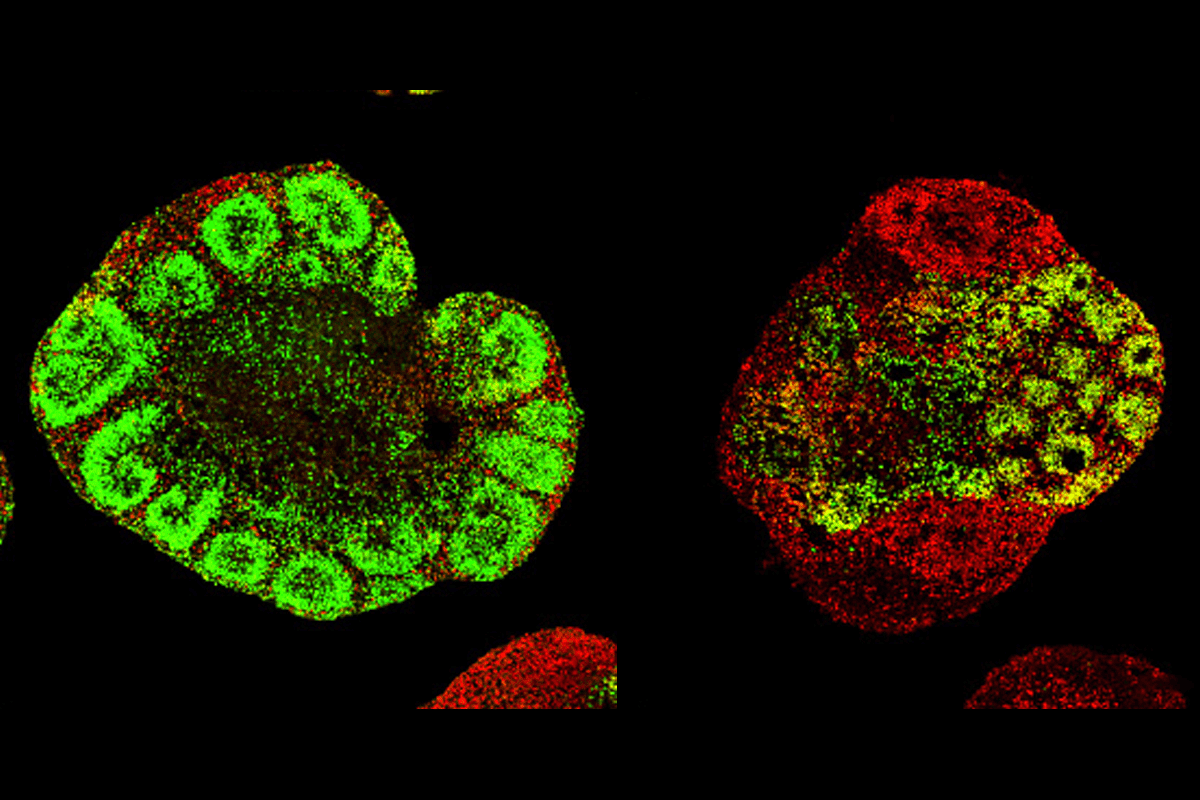
A simple protocol produces organoids that display molecular features of the front or rear of the human cortex, according to a new study. The technique could provide better cell-based models of autism and other neurological conditions that involve region-specific changes, the study suggests.
Briefly exposing neocortical organoids to signaling molecules called morphogens prompts them to develop their own signaling centers, the study found. These centers activate developmental pathways and induce molecular markers of anterior or posterior identity that match those found in the prenatal cortex.
“It’s a very significant step,” says Arnold Kriegstein, professor of neurology at the University of California, San Francisco, who was not involved in the work. The brain is more highly regionalized than any other organ, he says, with the same cell type taking on different functions and forming different connections depending on its location.
During embryonic development, gradients of morphogens organize the cortex along an anterior-posterior axis. But most cortical organoids either lack this large-scale organization or must be fused with separate, morphogen-secreting spheres to create assembloids.
The new technique instead stimulates organoids to form their own morphogen-secreting centers, avoiding the need to fuse tissues. It is “a very straightforward method” that is easy to replicate, says study investigator Momoko Watanabe, assistant professor of anatomy and neurobiology at the University of California, Irvine.
In the new study, Watanabe and her colleagues generated neocortical organoids from human pluripotent stem cells. After 15 days, the team exposed them to a fixed concentration of either FGF8 or BMP4, morphogens that promote anterior or posterior identity, respectively.
Organoids incubated with FGF8 for six days showed increased expression of anterior markers and formed FGF8-secreting clusters that signaled to nearby cells, helping to establish and maintain anterior identity. Similarly, organoids treated with BMP4 showed increased expression of posterior markers and developed posterior signaling centers that remained detectable at week 14, the study found.
The polarized organoids also showed shifts in gene expression along the anterior-posterior axis that matched those observed during fetal development.
T
But molecular markers are just one component of regionalization, Faravelli adds. “In the brain, areal identity also involves differences in cellular organization, connectivity and function, including interactions with other brain areas and the presence of sensory inputs. These organoids do not reproduce that full environment.”
Watanabe and her team also developed cortical organoids from stem cells derived from people with fragile X syndrome, a common genetic cause of autism.
Compared with control organoids, the fragile X models showed weaker gradients in the transcription factors SOX4 and SOX11 along the anterior-posterior axis. These findings mirror transcriptional changes that follow an anterior-posterior gradient detected in a previous analysis of cortical tissue from people with autism, highlighting the organoids’ potential to study region-specific changes linked to neurodevelopmental conditions.
The findings were published this month in Cell Stem Cell.
In addition to modeling neurological conditions, the organoids could help resolve the protomap-protocortex debate—that is, the extent to which cortical organization is preprogrammed or driven by external factors, such as incoming signals from the thalamus. The fact that cortical polarity can emerge without external input suggests that intrinsic programs initiate regionalization, Watanabe says. But refining those regions likely requires external inputs, which become increasingly important after birth, she says.
Watanabe and her team next plan to analyze the organoids beyond week 14, a stage that roughly corresponds to the third trimester of fetal development. By then, they expect to see more distinct patterns of molecular markers corresponding to more mature cortical organization, she says. In the long term, they would also like to investigate whether neuronal activity—measured using electrophysiology or calcium imaging—differs between anterior and posterior cortical organoids, she says.