New method creates uniform cultures of lab-grown neurons
A new technique leads to neuron cultures of consistent quality, enabling scientists to study how autism mutations alter neurons.
A new technique leads to neuron cultures of consistent quality, enabling scientists to study how autism mutations alter neurons1.
Mutations in genes linked to autism, such as SHANK2 and SHANK3, may lead to abnormal synapses — the junctions between neurons — and disrupted neuronal signaling.
Researchers often study the mutations’ effects in lab-grown neurons derived from a person’s skin or blood cells.
The method has problems, however: Not all the cells in a batch mature fully, so the number and density of neurons in a dish vary, as does the density of their connections2. This variability makes it difficult for scientists to spot differences between control neurons and those made from an autistic person.
In the new method, researchers controlled the density of neurons by growing a small number of new neurons atop a larger number of mature neurons of known density. They also cultured control neurons and neurons from an autistic person in the same dish, so that both developed under the same conditions.
Neuron lawn:
The researchers grew neurons made from two autistic children with SHANK2 mutations and from four controls; three of the controls were the children’s parents.
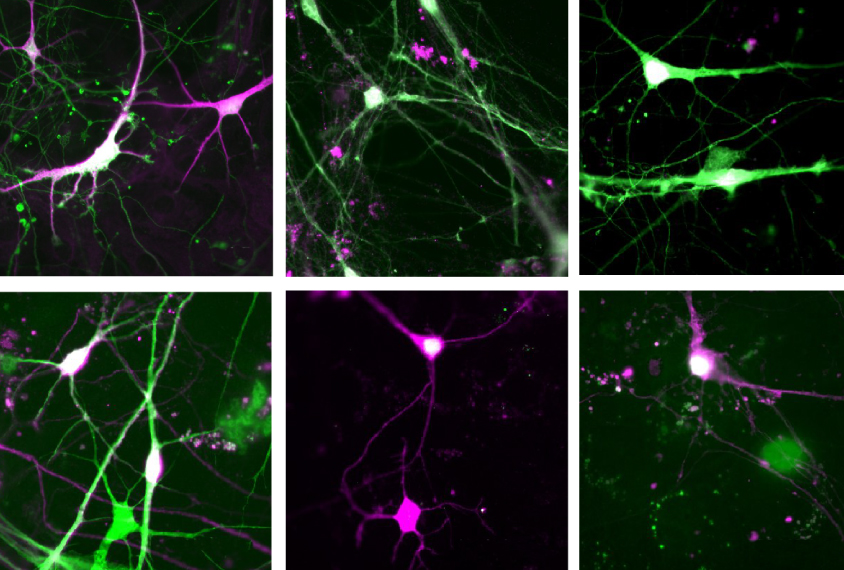
They tagged the autistic children’s neurons and control neurons with fluorescent proteins of different colors. They then transferred several thousand of both types of neurons to a dish lined with a lawn of about 200,000 control neurons and star-shaped support cells called astrocytes. After nine weeks, the researchers assessed the shape of the colored cells, the cells’ electrical activity and the number of synapses they had formed with other neurons.
The autistic children’s neurons have more synapses, more complex shapes, longer extensions and more electrical activity than do the control neurons, the researchers found. This finding contrasts with earlier work in rodent neurons suggesting that SHANK2 mutations decrease the number of connections between cells.
Researchers could use the new culture method, called SparCon, to study the effects of other autism genes on the form and function of neurons. They described the method in April in Nature Neuroscience.
References:
Recommended reading

Largest leucovorin-autism trial retracted
Pangenomic approaches to the genetics of autism, and more

Latest iteration of U.S. federal autism committee comes under fire
Explore more from The Transmitter
Cerebellum responds to language like cortical areas
Neuro’s ark: Understanding fast foraging with star-nosed moles
