This article is more than five years old.
Neuroscience—and science in general—is constantly evolving, so older articles may contain information or theories that have been reevaluated since their original publication date.
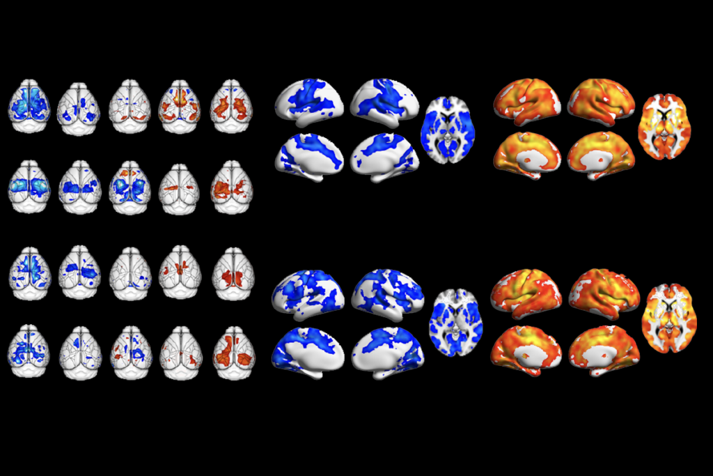
A collection of brain scans from monkeys aged 2 weeks to 12 months reveals how their brain structures and nerve tracts develop over time.
A collection of brain scans from infant monkeys reveals how their brain regions and nerve tracts develop over time1.
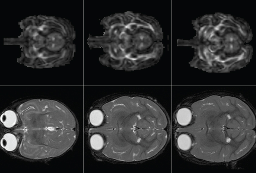
Existing atlases depict the brain structure of adult rhesus macaques. The new atlases, described 10 January in Frontiers in Neuroscience, contain structural brain scans for macaques aged 2 weeks to 18 months. The latter is the age equivalent of a 4- to 5-year-old child.
The collection could provide a reference point for researchers studying brain development in rhesus macaques, whose social behavior may yield clues about autism.
To create the atlases, researchers used structural magnetic resonance imaging (MRI) to visualize the brains of 40 rhesus macaques starting when the monkeys were 2 weeks old. The monkeys were anesthetized during the scans. The researchers repeated the scans at 3, 6, 12 and 18 months.
They used a custom-built device to hold the monkeys’ heads during the 75-minute scanning session. They first scanned various brain regions, such as the amygdala, which processes emotions, and the thalamus, a sensory relay center. Both of these regions are implicated in autism.
The researchers then used a technique called diffusion tensor imaging to capture the nerve tracts that connect brain regions. These tracts are called ‘white matter’ because of their pale color — a product of the fatty sheath that insulates each nerve fiber.
Macaques, like people, develop most of this insulation during infancy and early childhood — a process that may be altered in autism. For instance, a 2015 study showed that children with autism have atypical insulation in the corpus callosum, a white matter tract that bridges the brain’s hemispheres.
The atlases, which are available online, contain images from both types of brain scans, organized by age. Each image represents an average of all the monkeys scanned.