‘Mini-brains’ mirror fetal brain development
Spheres of brain cells derived from a fetal skull have gene expression patterns similar to those in brain tissue from the same fetus.
Spheres of brain cells derived from a fetal skull have gene expression patterns similar to those in brain tissue from the same fetus. Changes in their expression patterns over time also reflect those in the fetal brain.
Researchers presented the unpublished findings today at the 2017 Society for Neuroscience annual meeting in Washington, D.C.
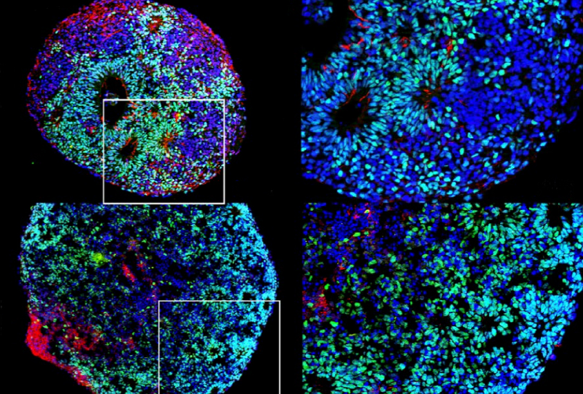
Scientists can reprogram stem cells derived from skin, blood or other tissues into clusters of neurons and other brain cells, called organoids, or ‘mini-brains.’ Depending on the cocktail of chemicals used to coax the cells along, the organoids can form layers of tissue resembling those in the brain.
Organoids are genetically identical to the cells they originate from, so researchers have been using them to study the effects of mutations associated with autism. But it is unclear how closely organoids mimic certain stages of brain development.
In the new work, researchers collected skull and brain samples from three fetuses between 15 and 17 weeks postconception. They used the bone cells to generate organoids, which they grew in the lab for more than 30 days.
The organoids closely recapitulate the fetal brain between 8 and 16 weeks postconception or earlier. “They are very close to the stage that we are trying to mimic [in autism studies],” says Soraya Scuderi, a postdoctoral associate in Flora Vaccarino’s lab at Yale University, who presented the work.
The work is part of the PsychENCODE project, which aims to characterize the role of noncoding DNA and chemical tags that alter gene expression in conditions such as autism.
Tracing timelines:
Scuderi and her colleagues also looked at gene expression patterns over time. They found that 2,680 genes increase or decrease in expression during the first 11 days of organoid development; another 643 genes ramp up or tamp down between 11 and 30 days in culture.
Genes that are expressed at high levels over the first 11 days and then fall are involved in cell division and DNA repair, whereas those that start low and then increase play a role in neuron outgrowth and signaling. This aligns with gene expression changes seen during the first 16 weeks of fetal brain development.
The findings support the use of organoids for studying stages of brain development that are relevant to autism, Scuderi says. “In the fetal brain, you can look at only one time point. In our organoids, we can look across time.”
The researchers also found changes over time in the activity of factors that regulate gene expression. These could be driving some of the patterns seen in the organoids, Scuderi says. She plans to use organoids to investigate how mutations in genomic regions that control gene expression may contribute to autism.
For more reports from the 2017 Society for Neuroscience annual meeting, please click here.
Recommended reading

New organoid atlas unveils four neurodevelopmental signatures

Glutamate receptors, mRNA transcripts and SYNGAP1; and more

Among brain changes studied in autism, spotlight shifts to subcortex
Explore more from The Transmitter

Not playing around: Why neuroscience needs toy models
