Pregnant rats deprived of oxygen for short periods during sleep — mimicking a disorder in people called sleep apnea — have an increased likelihood of giving birth to pups with an excess of synapses and autism-like traits, according to a new study.
Women who have sleep apnea periodically stop breathing while they sleep. Their male children are more likely than others to have altered neurodevelopment, according to post-hoc analyses in a small 2017 study. The new work is the first to test in an animal model how periodic oxygen restriction during pregnancy, or gestational intermittent hypoxia, might affect a developing child’s brain and behavior.
“Overall, gestational intermittent hypoxia appears to be a novel and translationally relevant animal model,” says Amanda Kentner, professor of psychology at the Massachusetts College of Pharmacy and Health Sciences in Boston, who was not involved in the work.
Fetal rats whose mothers experienced short periods of low oxygen during sleep did not show signs of oxygen deprivation themselves, the researchers found. But after birth, male offspring had altered communication, impaired cognitive function and atypical social behaviors — unlike their female littermates and control rats. The male offspring also had excess synapses in the cortex, the team found, a trait previously seen in mouse models of autism and in autistic people.
The work should encourage clinicians to consider screening pregnant people for sleep apnea, says lead investigator Michael Cahill, assistant professor of comparative biosciences at the University of Wisconsin-Madison. “Trying to go from rats to humans does have its challenges — I get that,” Cahill says. “But I think it’s something that should be considered more thoroughly.”
C
ahill and his colleagues placed pregnant rats into specialized chambers for sleeping from day 10 to 21 of their pregnancy. For one group, the chamber periodically limited the animals’ oxygen levels to model the nightly oxygen deprivation experienced by people with sleep apnea; for the other group, which served as a control, oxygen levels in the chamber remained normal.The treatment decreased blood oxygen levels in the pregnant rats, but not in their placentas or in their pups’ developing brains, the team found. However, pups born to rats that had experienced intermittent oxygen deprivation during sleep produced more ultrasonic vocalizations than those born to controls, Cahill and his colleagues found. When tested between 4 and 7 weeks of age, the males were less social and more cognitively impaired than either their female counterparts or controls of either sex.
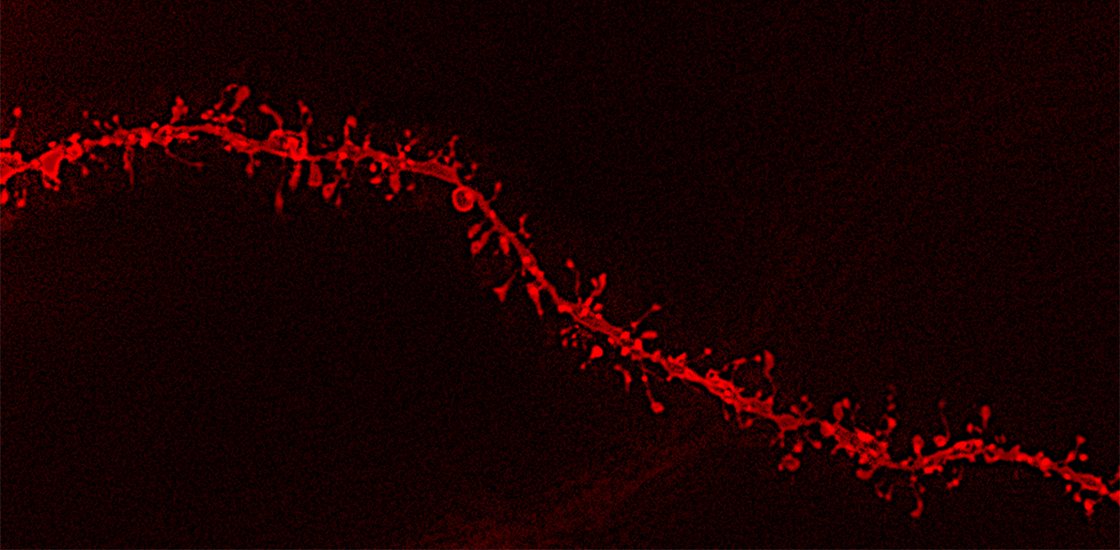
And at 8 weeks of age, the team found, those males had an atypically high density of dendritic spines — the nubs at which neurons receive signals from other cells — in their medial prefrontal cortex, a brain area that contributes to both social and cognitive functions. The findings were published in PLOS Biology in February.
The excess of synapses “is a very unusual phenotype in an animal model, but it’s actually something that occurs in autism,” Cahill says. It can stem from overactivation of the mTOR pathway, which regulates neuron growth and the formation of new dendritic spines.
In fact, the mTOR pathway is overactive in the offspring of rats that model sleep apnea, Cahill and his colleagues discovered. Implanting a pellet that steadily administers rapamycin, a drug that inhibits mTOR activity, prevented those animals from developing atypical behaviors.
The results add to a growing body of evidence supporting the idea that mTOR-related increased synaptic density “is a possible determinant of autism-related behavioral alterations,” says Alessandro Gozzi, senior researcher at the Istituto Italiano di Tecnologia in Rovereto, Italy, who was not involved in the new work.
T
he mechanism by which maternal sleep apnea affects the mTOR pathway, and thus neurodevelopment, remains unclear.Reduced oxygen levels can cause inflammation, according to previous research, and studies increasingly suggest that prenatal exposure to maternal inflammation can boost a child’s chances of having autism.
It’s also possible that the intermittent hypoxia treatment affects the animals’ sleep, Kentner says. Because sleep deprivation is also linked to inflammation, “I am not sure if it is the apnea in isolation or the combination of apnea and sleep deprivation from waking up — a two-hit model of sorts, if you will — that could be contributing to the offspring outcomes,” she says.
Kentner cautions against making strong claims about the sex differences the researchers observed. The team did not study the female rats into adulthood, meaning an atypical phenotype may just be delayed for the females, she says. And just because the male rats performed worse on the subset of tasks the researchers selected does not necessarily mean they would perform worse than female rats on all tasks, Kentner says.
Cahill agrees that the study may not fully capture the effects of gestational intermittent hypoxia in female rats. “Maybe we weren’t looking at the right behaviors to assess the female offspring,” he says.
In addition to investigating the potential sex differences, he and his colleagues say they plan to study whether blocking inflammation in the pregnant rats can prevent neuronal and behavioral changes in the offspring.


