Gerry Shaw / Wikimedia
Map of microglia in mouse brain may reveal role in autism
Researchers have mapped how immune cells in the brain called microglia change with age in mice.
Researchers have mapped how immune cells in the brain called microglia change with age in mice.
The findings pave the way toward a better understanding of these cells and their role in autism.
“Surprisingly, we still have no idea how and if they might contribute to [autism], mainly because we lack a basic knowledge about their biology,” says Timothy Hammond, a postdoctoral fellow in Beth Stevens’ lab at Boston Children’s Hospital. Hammond presented the unpublished findings today at the 2017 Society for Neuroscience annual meeting in Washington, D.C.
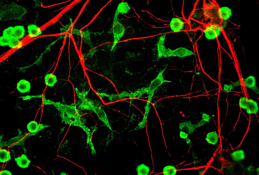
Microglia fight off pathogens and clear away debris such as incorrectly folded proteins. They are also thought to shape brain development by trimming the connections between neurons. In their inactive state, microglia typically assume a spider-like shape, but they morph into large blobs when they are actively degrading and engulfing substances.
Brain scans and multiple analyses of postmortem tissue suggest that microglia are excessively active in people with autism. It is unclear whether microglia directly contribute to autism or merely respond to changes in the brain that cause the condition, however.
Microglia ‘activation’ is a catchall term used for any microglia whose appearance deviates from the inactive state. But for these cells to be able to carry out so many diverse functions, there must be many different types of activated microglia.
“I think it’s well agreed upon in the field that there’s probably a diversity of activation states that they can assume,” Hammond says. “But we lack markers to really tease apart the functional differences between activation states and what these activation states mean in terms of how they’re influencing tissue.”
Microglia galore:
Hammond and his colleagues explored the diversity of microglia by isolating more than 75,000 of the cells from mice and analyzing each one. They isolated cells from mouse embryos and from mice at 5, 30 and 100 days old.
They used a relatively new method to measure the levels of genes that individual microglia express. A computer program then grouped the cells based on similarities in their gene expression patterns.
This analysis revealed 11 types of microglia; 7 of the types mostly consist of cells from embryonic and newborn mice, and 1 cluster is made up of only cells from juvenile and adult mice.
“The microglia diversity really seems to be the greatest early on in development,” Hammond says.
All 11 groups express genes already known to be characteristic of microglia. But each group also expresses a unique set of genes. The researchers used these ‘genetic fingerprints’ to map where each of the 11 microglia cell types reside in the brain.
For example, one type of microglia uniquely expresses the SPP1 gene. Using brain slices from newborn mice, the researchers found that the SPP1 subtype of microglia reside only in the cerebellum and developing nerve bundles.
“This could potentially allow us to use these molecular signatures we found in each of these subsets to start to dissect apart new and interesting functions for these cells,” Hammond says.
The researchers are using these signatures to map the locations of microglia in postmortem brain tissue from people with autism as well as from mice with mutations in SHANK3 or CHD8, two top autism risk genes. They are also mining gene expression datasets to see whether the signatures for the 11 microglia subtypes are altered in autism.
For more reports from the 2017 Society for Neuroscience annual meeting, please click here.
Recommended reading
Explore more from The Transmitter

This paper changed my life: John Tuthill reflects on the subjectivity of selfhood

Some facial expressions are less reflexive than previously thought


