Map of gene expression gives new clues to brain development
A new map that analyzes gene expression one cell at a time shows how various cell types mature and form the brain’s distinctive structures.
A new map that analyzes gene expression one cell at a time shows how various cell types mature and form the brain’s distinctive structures1. The findings suggest that a particular type of neural stem cell plays a role in autism.
During development, cells called glia generate new neurons. These neurons mature into distinct types and migrate to what will become the six layers of the brain’s outer shell, the cerebral cortex. This process is critical to establishing mature neuronal circuits.
The researchers isolated single cells from 48 tissue samples taken from 9 brain regions in a human embryo or fetus. The samples spanned 29 time points ranging from just shy of 6 weeks to 37 weeks after conception. The researchers isolated more than 4,000 individual cells and sequenced the RNA of each to measure its gene expression.
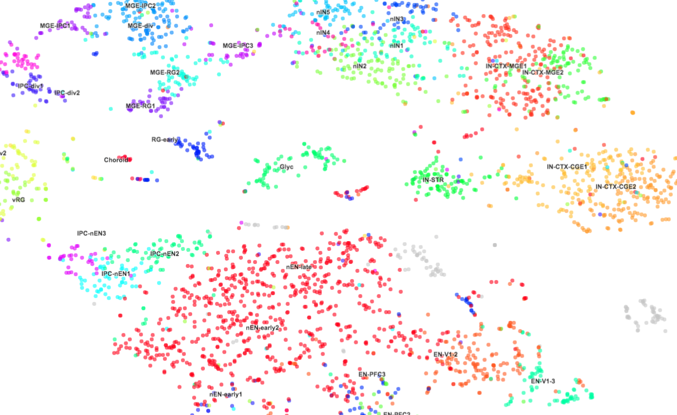
Using a statistical analysis of the gene expression patterns, the researchers identified 11 categories of cells and analyzed how the cells change during development.
Radial glia cells — a type of stem cell that gives rise to new neurons — mature faster in the prefrontal cortex (which governs judgment and planning) than in the visual cortex. The researchers also found expression patterns that separate stem cells destined to form deep brain structures from those that form its outer layers.
Specific gene expression patterns established at about 15.5 weeks after conception help define cortical layers, they found.
The researchers also checked for cells expressing high levels of 65 top autism risk genes. They found that neural stem cells called ‘outer radial glia’ express autism genes in a cell signaling pathway called mTOR. This expression occurs during the period when most neurons are born.
Outer radial glia cells may be especially sensitive to mutations affecting the mTOR pathway, the researchers say. This vulnerability could help explain changes that underlie autism.
The researchers compiled the results in an open-source online map called the UCSC Cluster Browser. The complete dataset can be explored by brain area, layer, cell type, sample name and age.
References:
- Nowakowski T.J. et al. Science 358, 1318-1323 (2017) PubMed
Recommended reading
New tool may help untangle downstream effects of autism-linked genes
NIH neurodevelopmental assessment system now available as iPad app

Molecular changes after MECP2 loss may drive Rett syndrome traits
Explore more from The Transmitter
Organoids and assembloids offer a new window into human brain
Who funds your basic neuroscience research? Help The Transmitter compile a list of funding sources
