Interactive mouse brain atlas helps researchers share data
An online software suite charts individual neurons and their connections across the mouse brain.
A software suite charts individual neurons and their connections across the mouse brain via a web-based platform1. The maps could help scientists study neuronal connections in mouse models of autism.
Several large-scale efforts have mapped mouse brain-cell types and the connections among them. But each one organizes, visualizes and processes data differently, making efforts to translate them into sharable digital maps laborious.
The new platform allows scientists to upload their mouse brain-imaging data and visualize individual cells, their connections and gene expression patterns in a standardized atlas. Scientists then can easily share data and results with collaborators.
The software can work with images captured via widefield, confocal or light-sheet fluorescent microscopy, and does not require programming experience to use.
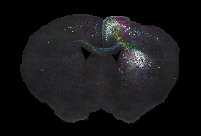
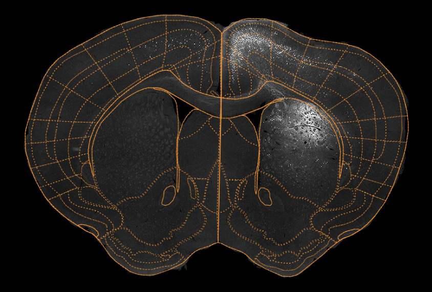
It can layer different types of information, so scientists can simultaneously compare cell types, the connections between neurons and neuronal activity (as revealed by gene expression patterns in the tissue). It can also locate features in imaging data, such as cell bodies and fiber tracts, by mapping them to coordinates in the Allen Institute’s Mouse Brain Atlas.

Scientists can visualize these matched data as a graphic overlay on their original images or on a digital version of the reference brain. The researchers described the tool 4 December in Nature Neuroscience.
To demonstrate the software, the researchers used data from mice given an injection of cocaine. Brain sections from the mice revealed increased neuronal activity in an area called the orbitofrontal cortex, which integrates sensory information and is involved in decision-making.
Adding the images to the reference atlas reveals connections between the orbital cortex and two other regions: the primary motor cortex, which controls movement, and the striatum, part of the brain’s reward system. The connections make up a circuit that mediates cocaine’s effects, including the ‘high’ and increased movement.
References:
- Fürth D. et al. Nat. Neurosci. 21, 139-149 (2018) PubMed
Recommended reading

Expediting clinical trials for profound autism: Q&A with Matthew State

Too much or too little brain synchrony may underlie autism subtypes
Explore more from The Transmitter

Mitochondrial ‘landscape’ shifts across human brain

