As animals carry out complex behaviors, multiple brain areas turn on and talk to one another. But neuroscientists have had limited means to measure that neuronal dialogue. Electrical recordings, for example, are typically constrained to one brain area at a time, or require that mice have their head fixed in a specific position.
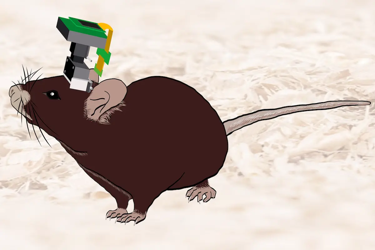
A new technology overcomes those restrictions. The device, called E-Scope, reported in a peer-reviewed preprint in eLife, effectively measures the activity of neurons in two different areas at the same time, even as rodents move freely.
The headset captures images of calcium currents, made using a microscope, and recordings of neurons’ electrical activity through electrodes to show how the cerebellum communicates with other brain regions during social interaction in mice. “Everything [is] synchronized together that way,” says Peyman Golshani, assistant professor of neurology at the University of California, Los Angeles and a study investigator.
This approach holds the potential to illuminate how coordination between brain areas in conditions marked by impaired social interaction, such as attention-deficit/hyperactivity disorder and autism, is disrupted, Golshani says. By combining technologies, researchers who use the E-Scope “don’t need separate electrophysiology and imaging hardware,” he adds.
It’s also much more comfortable for the animals, according to Golshani. A single wire conveys all of the small headset’s data, so mice can move more freely than when wearing other devices.
U
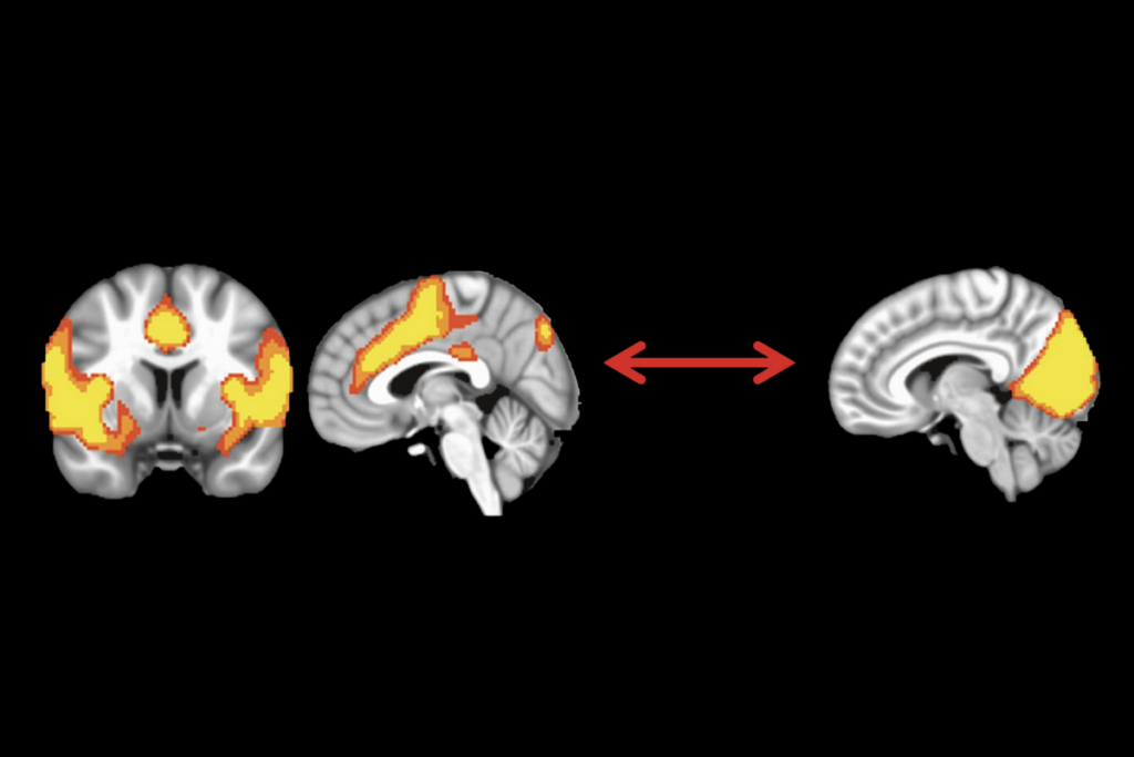
sing the E-Scope, the group studied activity in two distant but interconnected regions: the cerebellum and the anterior cingulate cortex (ACC). Disrupting these two areas makes animals less sociable, according to previous studies, so Golshani and his team set out to examine how they talk to one another while mice socialize.Wildtype mice wearing the E-Scope interacted with either an object or another mouse for seven minutes. During social interactions, neurons in an area of the cerebellum called the dentate nucleus fired, while brain cells called Purkinje neurons became silent.
The changes in the cerebellum occurred when the mice were interacting with each other but not during other behaviors, such as head movements. According to one peer reviewer who commented on the preprint, these observations begin to reveal the non-motor functions of the cerebellum, which are understudied.
Similarly, within the ACC, some neurons engaged only during social interactions. And changes in the ACC and cerebellum during socialization correlated with each other more strongly when mice interacted with each other but not with an object, suggesting that the connections between them could be coordinating social behavior.
The findings demonstrate the E-Scope’s abilities—but, as the reviewer noted, further investigation is needed to understand how the interacting activity between the ACC and cerebellum relate to social behavior. The data coming from the two brain regions are only correlational, that reviewer wrote, and so “the causal relationship is far from established.”
One challenge the E-Scope presents for users is accessing enough brain area or “real estate,” as Golshani calls it, to obtain significant data, a limitation he and his colleagues encountered when recording calcium channels in the cerebellum.
“Sometimes we would just get a few neurons per animal, maybe one or two,” Golshani says. Plus, the device is still quite bulky and heavy for mice and could be miniaturized even further, he adds.
Details and tutorials for building the device are open access, so others can develop their own versions and perform novel studies. Although Golshani and his group are interested in how other brains areas, such as the nucleus accumbens, modulate social interaction in models of autism, he points out the E-Scope can also “be used for any other type of behavior.”


