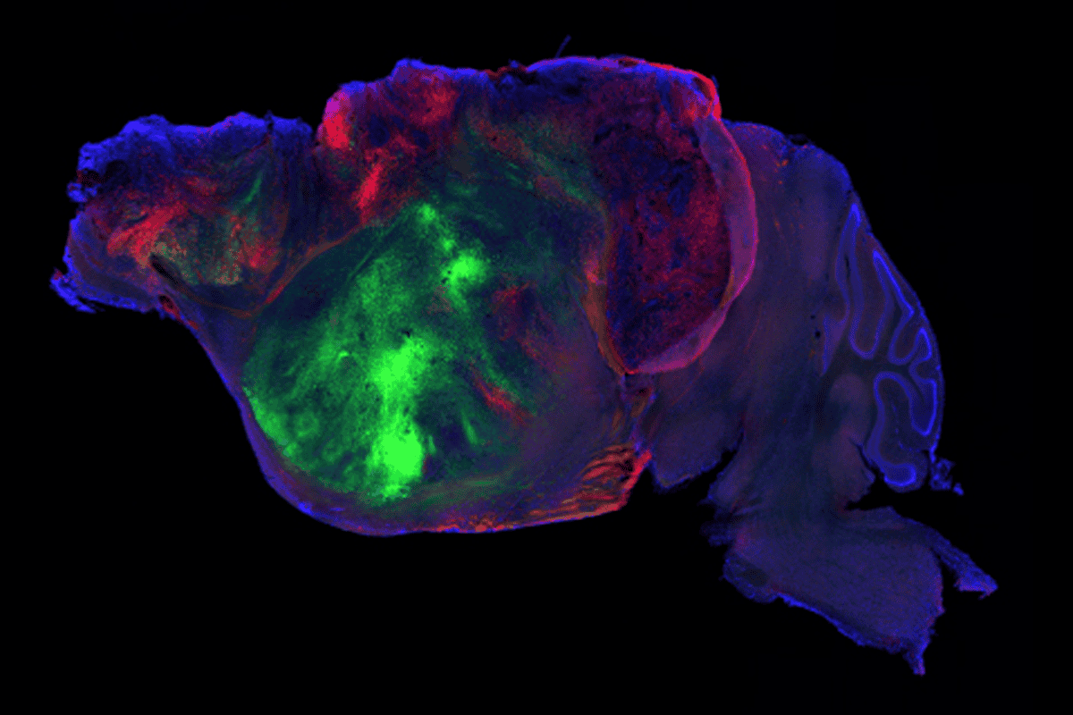
Human cortical organoids transplanted into newborn mice integrate into the nervous system and form circuits that support electrical activity, according to a new study published today in Nature. The mice were engineered to lack nearly all of their cerebral cortex, creating space for the human-derived tissue to take hold.
The researchers then used the ‘xenocortical’ mice to model hypoxia, a state that occurs when brain cells don’t get enough oxygen. When hypoxia occurs prenatally or around the time of birth, it can cause cerebral palsy; it is also associated with autism and epilepsy.
The transplantation approach could serve as a new tool to study typical development and neurodevelopmental conditions, as well as test potential therapeutics, says Hongjun Song, professor of neuroscience at the University of Pennsylvania’s Perlman School of Medicine, who was not involved in the study.
“Now you have a model, a live model where you can actually test all of this with human cells in live animals,” Song says.
The lack of access to living brain tissue has presented a long-standing barrier to understanding human development. Brain organoids derived from human stem cells have opened up fresh possibilities, and transplanting the organoids into animals such as rats can help the human cells mature even further and form more complex circuits. But physical space within the rodents is finite, and the human neurons can struggle to establish themselves.
“They will be outcompeted by mouse cells very quickly,” says Sergiu Pașca, professor of psychiatry and behavioral sciences at Stanford University and the study’s senior investigator. “It’s also very difficult to actually distentangle, to be honest, the effect of the human versus the mouse.”
In the new paper, Pașca and his team used a knockout strategy to design a mouse model. They deleted ESCO2, a protein that aids in cell division, from cells expressing a marker for the dorsal and medial pallium, the areas of the embryonic forebrain that give rise to the cortex and hippocampus. The team also designed the mice to be immunocompromised so that the animals would not reject the human cells.
The resulting mice were missing 98 percent of their cortex and hippocampus and had a 50 percent decrease in total brain tissue, compared with controls. Even so, the mice exhibited only minor behavioral changes, such as having a more cautious gait.
“These animals are not perfectly normal but also surprisingly functional, much more than we thought they would be,” Pașca says.