Homemade ‘Miniscope’ lights up firing neurons in mobile mice
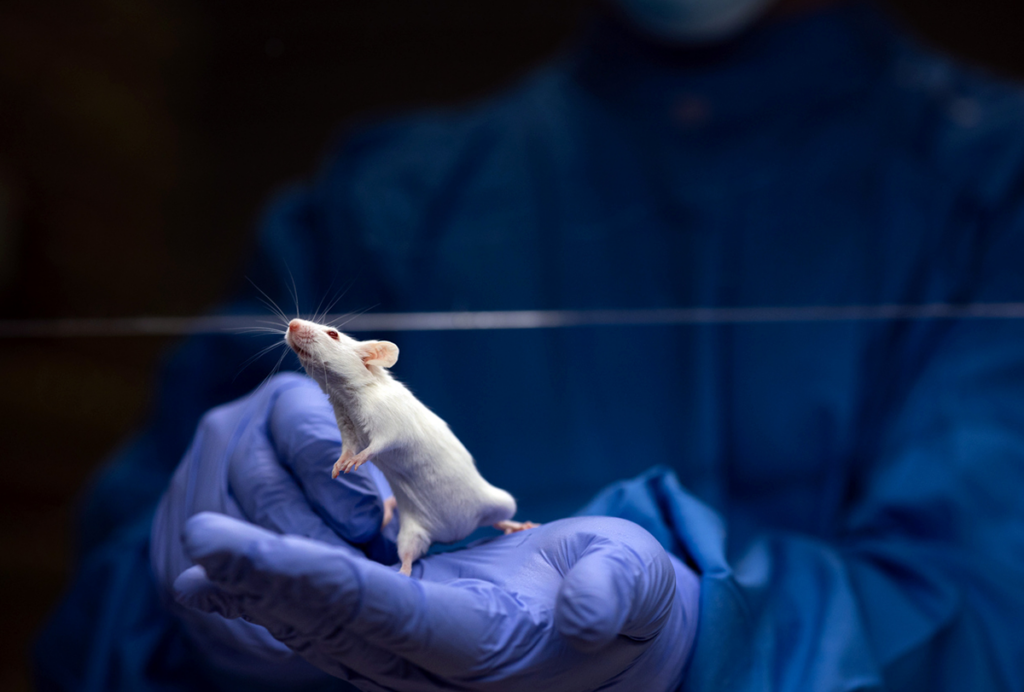
A miniature microscope made from cheap, ‘off-the-shelf’ parts can track firing neurons in the brains of freely moving mice.
A miniature microscope made from cheap, ‘off-the-shelf’ parts can track firing neurons in the brains of freely moving mice. Researchers presented the blueprints for building the ‘Miniscope’ today at the 2015 Society for Neuroscience annual meeting in Chicago.
Existing head-mounted microscope systems can cost upward of $150,000, which includes the cost of two microscopes and a readout system. By contrast, researchers can build five Miniscopes with a readout system for roughly $3,000.
Given the potential applications of the scope, word has travelled fast. “We’ve been contacted by a lot of postdocs who are leaving labs and starting their own, and they don’t want to spend all their start-up funds on one system,” says Daniel Aharoni, a postdoctoral fellow in Peyman Golshani’s lab at the University of California, Los Angeles (UCLA), who presented the work. “This is a great option for them.”
The Miniscope is a fluorescent microscope, roughly the height of a Lego brick, that weighs less than 3 grams. It snaps via a magnet onto a baseplate implanted in a mouse’s head — akin to a MacBook charger. This allows researchers to easily connect the microscope for experiments without needing to anesthetize the mouse each time.
Researchers can adjust the height of the Miniscope’s imaging sensor to visualize neurons at various depths. But because the device is so cheap, they recommend using the same Miniscope at the same depth throughout an experiment.
“That way, every time you go back with the microscope, you’re hitting the same focal plane,” says Aharoni.
Pint-size power:
A slim wire connects the microscope to a power source and software for analyzing the data. The wire is “basically weightless,” according to Aharoni, and less than one-third of a millimeter thick, allowing mice to roam their cages unencumbered.
To test the microscope, the researchers first injected a virus into the mouse hippocampus, a brain area involved in learning and memory. Under fluorescent light, the virus illuminates cells that are flooded with calcium, which triggers neurons to fire.
The researchers then implanted a tiny lens just above the hippocampus and cemented it in place with a baseplate. When the microscope is attached to the baseplate, it shines fluorescent light on the neurons, revealing which ones are firing. A miniscule camera records the firing patterns in real time.
The images are detailed enough to distinguish every neuron from its neighbor, but not so detailed that the files are unwieldy.
The system can also record behavior in the mice by tracking the position of a red light mounted on top of the microscope. This feature could help autism researchers track the activity of specific neurons during social interactions.
“We can then see how these cells malfunction in mouse models of autism,” says Golshani, associate professor of neurology at UCLA’s David Geffen School of Medicine. “I think this will be really critical for understanding which circuits are malfunctioning, and for testing potential treatments to see if they correct the functional disturbance.”
The researchers are already developing a wireless version of the Miniscope and a version that integrates optogenetics — a technique that uses blue light to activate specific neurons. They plan to post instructions for how to build the devices at miniscope.org and are offering classes at UCLA and online tutorials to help other researchers use them.
For more reports from the 2015 Society for Neuroscience annual meeting, please click here.
Recommended reading

New organoid atlas unveils four neurodevelopmental signatures

Glutamate receptors, mRNA transcripts and SYNGAP1; and more

Among brain changes studied in autism, spotlight shifts to subcortex
Explore more from The Transmitter

Psychedelics research in rodents has a behavior problem
Can neuroscientists decode memories solely from a map of synaptic connections?
