Several proteins with strong links to autism are also tied to cilia, the hair-like structures on cells, a new preprint finds. Variants in the genes that encode these proteins may relate to co-occurring cilia-related conditions that often affect the quality of life for autistic people as well, the study investigators say.
Fragile X syndrome, tuberous sclerosis complex, focal cortical dysplasia, CDKL5 deficiency disorder and Rett syndrome may also be linked to cilia, says Hye Young Lee, associate professor of cellular and integrative physiology at the University of Texas Health Science Center at San Antonio, who was not involved in this study. The new findings therefore suggest “very interesting future works for many cilia or autism researchers,” she says.
Multiple autism-linked genes converge around the function of cellular structures called microtubules—particularly those that make up cilia, the study investigators previously found. Most cells of the body have primary cilia, which often serve sensory or signaling functions. Some cells, including those lining the airways, also have motile cilia, which help sweep fluids such as mucus, among other functions.
Several autism-linked variants are tied to cilia defects, research from the past decade suggests. And autism often co-occurs with cilia-related disorders, or ciliopathies, such as congenital heart disease, slow-moving gut, epilepsy and respiratory issues, “especially when it comes to rare variants of autism-linked genes,” says preprint investigator Elina Kostyanovskaya, a doctoral candidate in Helen Willsey’s lab at the University of California, San Francisco.
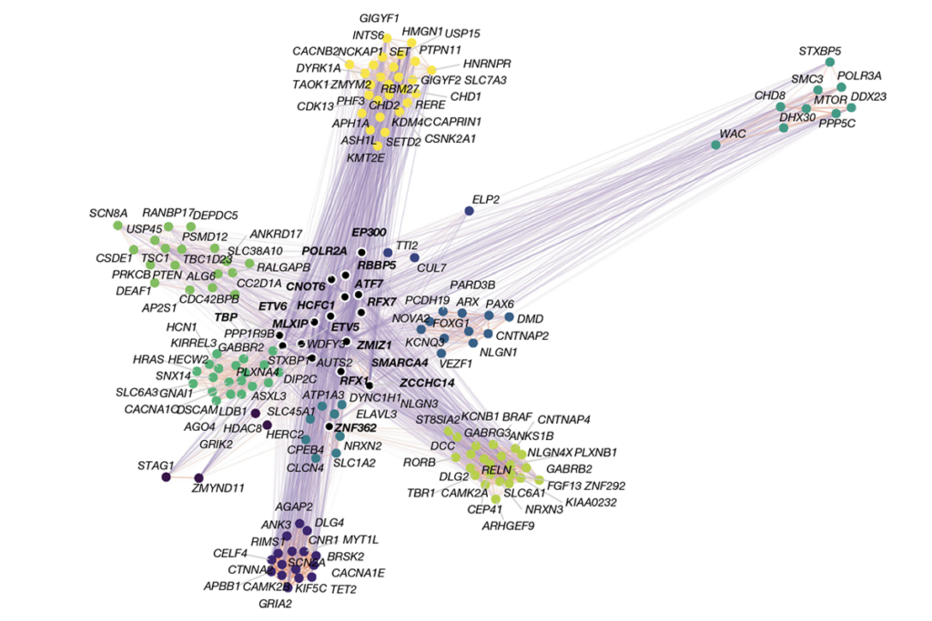
In the new study, Kostyanovskaya, Willsey and their colleagues analyzed 255 genes that had been strongly linked to autism in a 2022 meta-analysis and found that the expression of 101 of these genes in human neurons, glial cells and other brain cells tracks with that of ARL13B, a key marker of primary cilia.
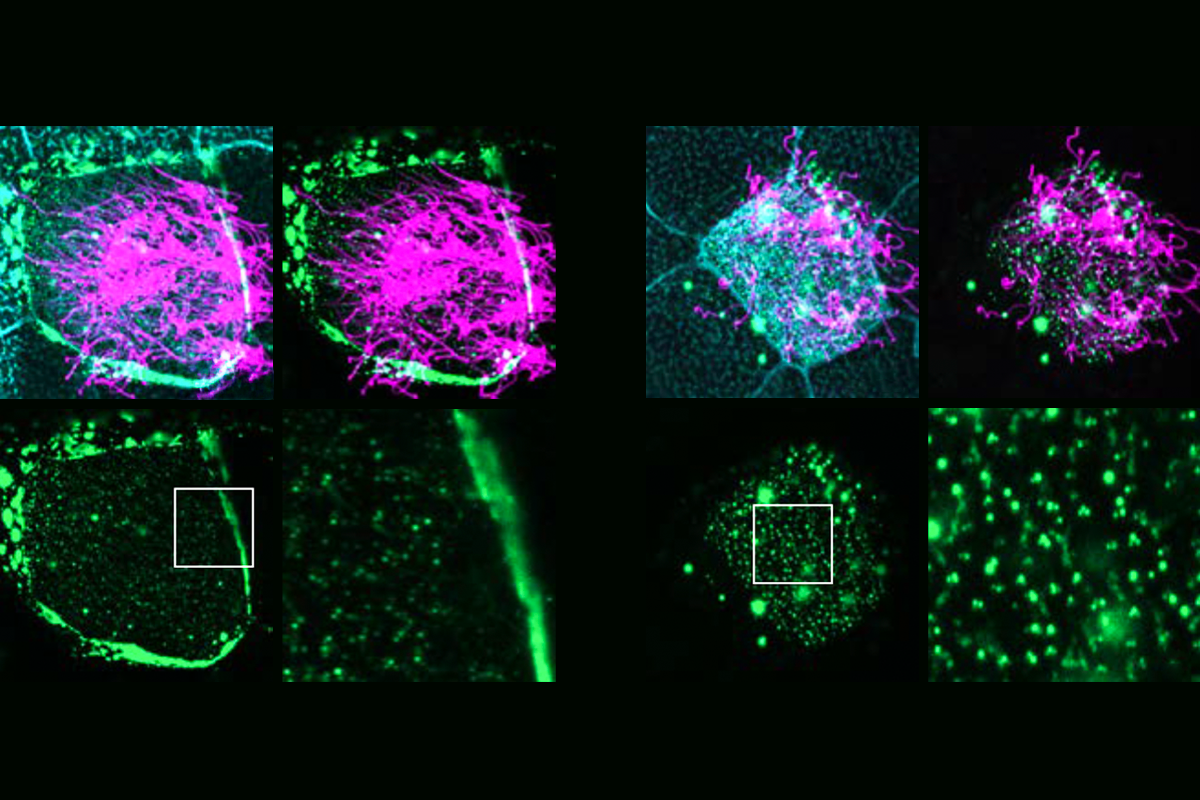
The proteins encoded by 12 of these genes are located in or near primary cilia in rat neurons and motile cilia in frog epidermal cells, the scientists discovered by tagging 30 of the top genes with green fluorescent protein. These proteins were previously categorized as gene-expression regulators or as involved in neuronal communication.
“The dogma is that genes strongly associated with autism predominantly function either at the synapse for neuronal communication or at chromatin for gene expression regulation,” Kostyanovskaya says. “We’re saying that even if a gene has one well-established function, it may have more.”
O
ne of the proteins examined, SYNGAP1, is thought to support learning and memory at synapses. This protein is present in the cilia of frog, mouse, rat and human cells and is necessary for cilia formation in frogs, the researchers found. But it concentrates away from frog cilia, either at the plasma membrane or inside the cytoplasm, when it carries either of two variants derived from autistic people. The findings appeared in a preprint the team posted 5 December on bioRxiv.Ultimately, these findings “could lead to novel biomarkers for earlier and more accurate diagnoses” of autism, says Thomas Theil, reader in developmental biology at the University of Edinburgh, who was not involved in this research.
For instance, people with cilia defects generate unusually low levels of nitric oxide gas from their nasal ciliated cells. Kostyanovskaya and her colleagues found that a 30-second test with a nasal probe detected significantly low nasal nitric oxide levels in 21 of 24 people with SYNGAP1 variants when compared with unaffected family members. “If we can measure people with this issue related to autism in this pretty noninvasive manner pretty quickly, that’s exciting,” she says.
Still, “many more studies will need to be done” to prove that cilia defects contribute to autism, says Mustafa Khokha, director of Guerin Children’s Genetics Research Center at Cedars-Sinai Medical Center, who was not involved in the new work.
For instance, he says, if a study deletes a protein that works at both the synapse and at the cilium, “how do you know that the phenotype is caused by effects at the synapse versus the cilium?” Experiments that can localize the protein to either the cilia or synapses are “definitely possible, but challenging.”
Future research should examine whether autism-linked proteins are involved in cilia-related processes such as cilia formation or signaling, says Kerstin Hasenpusch-Theil, a postdoctoral researcher in Theil’s lab. “It is also critical to gain a better understanding of whether and how dysregulated ciliary processes contribute to autism development,” Hasenpusch-Theil adds.
In the end, “are all types of autism going to have a ciliary component?” Kostyanovskaya says. “Probably not. But a large component may, especially those linked with rare gene variants linked with autism.”
