Compound lets scientists see deep inside brains of living mice
A chemical that doctors use to create contrast on X-rays also yields clear images of neurons in the brains of living mice.
A chemical that doctors use to create contrast on X-rays yields clear images of neurons in the brains of living mice, according to unpublished findings presented Tuesday at the 2017 Society for Neuroscience annual meeting in Washington, D.C.
Autism researchers could use the method to peer inside the brains of mouse models of the condition.
“Some of the ways people have investigated parts of the brain that they can’t access easily with light is they excavated parts of the brain or shove a lens into the brain,” says Maiko Kume, graduate student in Tim Holy’s lab at Washington University in St. Louis, who presented the work. “We think it would be a less invasive way to use light to investigate the brain.”
Researchers have traditionally created images of animal brains by slicing them into thin sections and freezing proteins or cells they aim to visualize. Then they wash away lipids and other occluding molecules to make the tissue transparent. But these clearing methods aren’t suitable for living animals.
Pictures of neurons in living mice often look blurry. When light passes through cells and the fluid that surrounds them, the image becomes distorted due to ‘refraction,’ or bending of the light. A similar distortion makes a pencil appear broken when partially submerged in water.
The new technique allows researchers to look deeper into the brain without running into this distortion. It involves a compound called iodixanol, which moderates the refraction of light as it passes through tissue.
Clear view:
Doctors inject iodixanol into people’s veins to create contrast between organs and blood vessels and, ultimately, to make certain tissues stand out in an X-ray. For instance, it’s commonly used to help detect blood clots.
Kume and her colleagues tried it on mouse brains. First they used mouse brain tissue, immersing brain slices in the compound for 30 minutes. The effect was obvious, even to the naked eye. The tissue was suddenly see-through, as paper becomes when water is spilled on it.
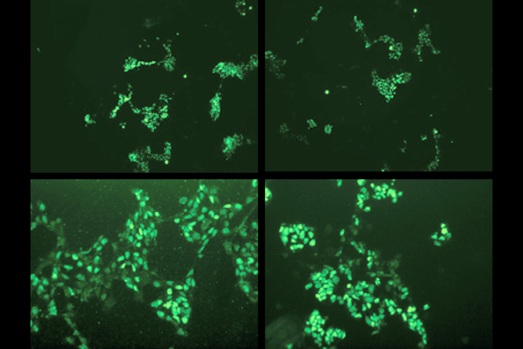
Under a microscope, the effect was also evident. Fluorescently labeled neurons were visible at much greater tissue depths when using iodixanol.
The researchers then tried the technique in living mice. They gave the mice anesthesia and surgically etched a window into their brains. After labeling their neurons with a fluorescent marker, they used a microscope to capture the mouse neurons without the compound. Then they injected the compound into the brain, waited for it to diffuse through the tissue, and took a picture of the neurons again.
The difference is striking. The first images show scattered patches of neurons. The second ones are alight with a starry sky of full of neurons, indicating that the picture extends deep into the tissue.
A day later, the researchers put the mice through behavioral tests to measure the effect of the infusion. They ate, drank and groomed themselves normally and performed balance and strength tests as well as mice that underwent surgery but weren’t infused with the compound. The treated mice were, however, less active and less apt to explore their environment than the other mice.
“We were not surprised that infusing a large amount of this compound into the brain will affect activity. We were happy that the mice survived,” Kume says.
As a next step, Kume plans to use calcium imaging in awake mice to get snapshots of their brain activity in real time.
For more reports from the 2017 Society for Neuroscience annual meeting, please click here.
Recommended reading
Developmental delay patterns differ with diagnosis; and more
Split gene therapy delivers promise in mice modeling Dravet syndrome
Changes in autism scores across childhood differ between girls and boys
Explore more from The Transmitter
Smell studies often use unnaturally high odor concentrations, analysis reveals

‘Natural Neuroscience: Toward a Systems Neuroscience of Natural Behaviors,’ an excerpt
