This article is more than five years old.
Neuroscience—and science in general—is constantly evolving, so older articles may contain information or theories that have been reevaluated since their original publication date.
A potent chemical cocktail renders tissue transparent in a way that makes even buried brain structures visible.
A potent chemical cocktail renders tissue transparent in a way that makes even buried brain structures visible. Researchers can use the recipe, detailed 22 August in Nature Methods, to glimpse neurons and other delicate structures inside the intact bodies of mice1.
Existing techniques, such as CLARITY, CUBIC and iDISCO, can make solid tissue see-through. Researchers have used these methods to visualize fluorescent proteins, which tag structures inside animals that have been genetically modified to carry these fluorescent tags. But the chemical ingredients of these tissue-clearing brews can dim the proteins’ glow.
Researchers have worked around this problem by adding the fluorescent tags after tissues have been made transparent. But the deeper these tags are, the harder they are to see.
The new method, dubbed uDISCO for ‘ultimate’ DISCO, uses a unique mix of chemicals that preserves more of the fluorescence in the protein tags. It also shrinks tissue samples, allowing scientists to see deeper into the tissue and view the entire body of a mouse at once.
In the study, researchers used uDISCO to visualize the complete nervous system of an adult mouse. They removed the skin of the mouse and drained its blood, which can give off a natural fluorescence. They then bathed the body in a series of chemical cocktails: one that removes water, another that dissolves fat and a third that makes tissues — even bones — transparent. The entire process takes three to four days.
The technique shrinks the body of an adult mouse by about 42 percent, bringing buried proteins closer to the surface. The small size of the specimens also allows researchers to study swaths of tissue up to four times larger than what they could visualize with existing clearing methods. This greater coverage makes even long-range connections traceable: The researchers were able to see the entire optic nerve, a bridge between the eyes and the brain.
The researchers also applied uDISCO to the dissected brains and spinal cords of mice, shrinking this tissue by about 55 percent to capture high-resolution images of deep neurons and their branches.
The researchers also mapped the paths of blood vessels in the brains and spinal cords of rats (see video above). This task has traditionally involved slicing tissue into ultra-thin sections, imaging each section, and aligning the images to construct a three-dimensional map of the vessels. Using uDISCO, the process was easier and more accurate — and the researchers were able to see bone marrow stem cells inside the vessels.
Scientists could use uDISCO to trace the connections between neurons in mouse models of autism or in postmortem brain tissue from people with the condition.
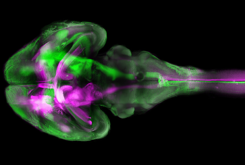
Color cascade: Different nerve tracts glow in green and purple in a transparent mouse brain.
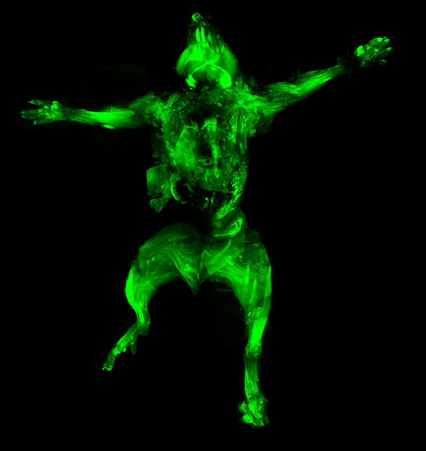
Full body: Researchers can use uDISCO to visualize the complete nervous system of a mouse.
See-through specimen: uDISCO renders a mouse brain transparent.
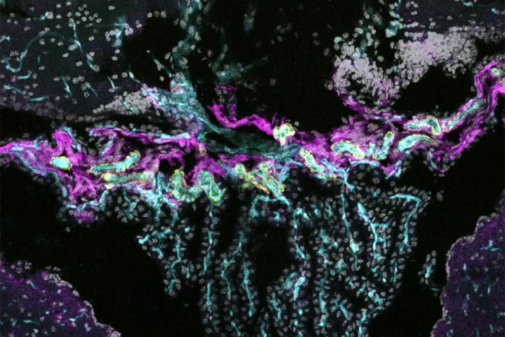
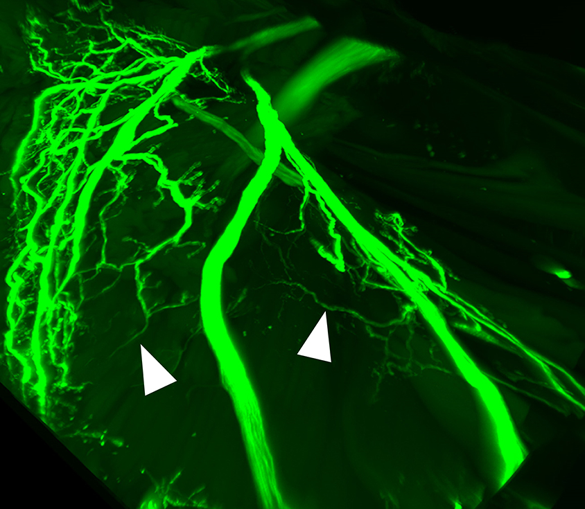
Close up: The method reveals the delicate branches of neurons connecting a mouse’s brain to its whiskers.
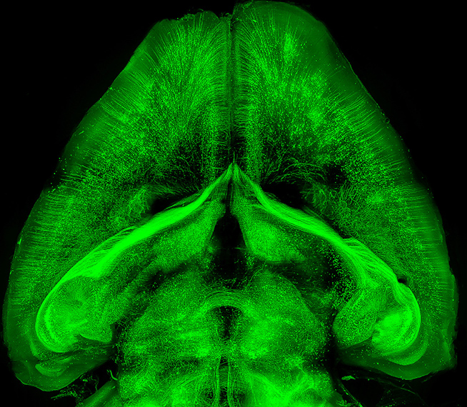
All in one: Applying uDISCO directly to a dissected mouse brain and spinal cord allows researchers to capture the expansive central nervous system in a single image.
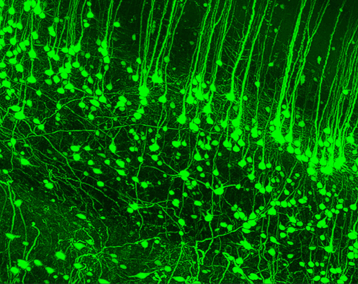
Fine detail: Neurons in the mouse’s cortex, the outer layer of the brain, glow brightly.
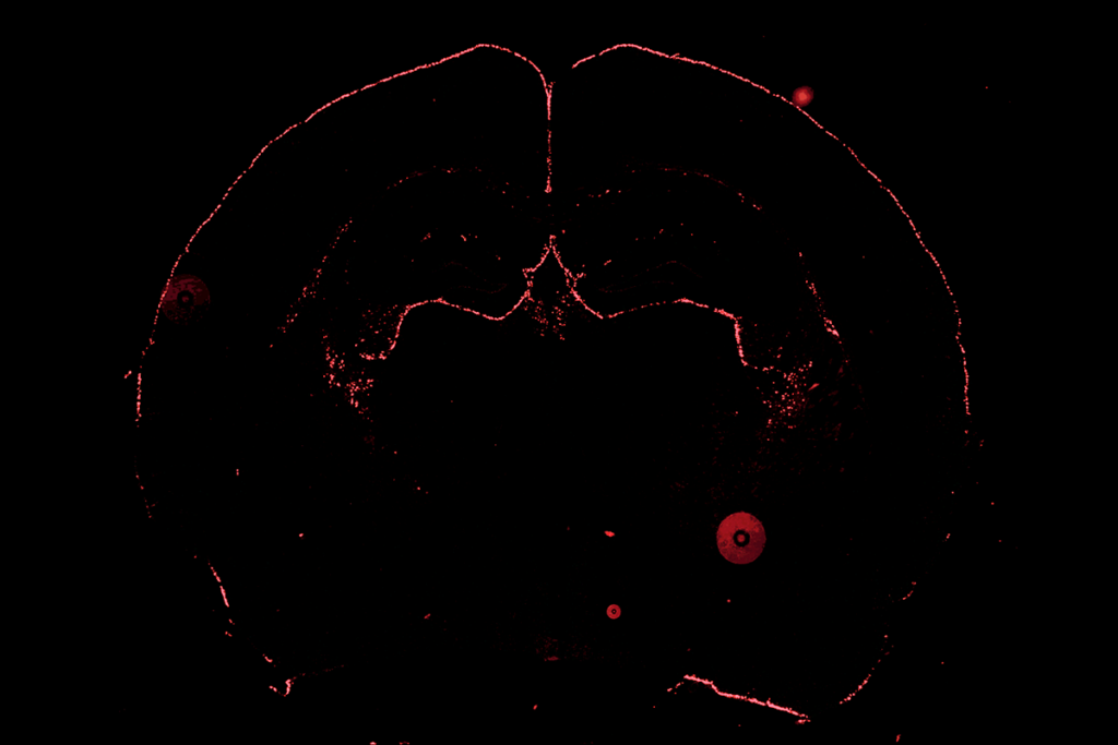
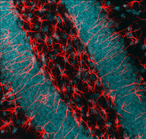
Sorting cells: Cleared brain tissue contains a mix of neurons (nuclei shown in blue) and star-shaped cells called astrocytes (red).