Brain scans reveal subtypes of fragile X syndrome in boys
Differences in brain structure may distinguish boys with relatively mild features of fragile X syndrome from those with a severe form of the condition.
Differences in brain structure distinguish boys with relatively mild features of fragile X syndrome from those with a severe form of the condition, a new study suggests1.
Enlarged brain regions — and big brains — mark the boys with significant intellectual disability and difficulty in everyday functioning. Boys without enlarged brains, by contrast, have milder features, the study reports.
Fragile X syndrome is an inherited form of intellectual disability; more than one-third of boys with the condition also have autism. Scientists have long suspected that the syndrome has subtypes because of the range of cognitive and other skills seen in these children. But no one had found a rigorous way to group people with the condition.
“This is really the first approach that seems to yield these different subgroups,” says lead investigator Allan Reiss, professor of psychiatry and behavioral sciences at Stanford University in California.
Identifying subtypes of fragile X syndrome may help scientists tailor therapies for individuals with the condition.
“If you can stratify using these kinds of neuroanatomical measures, you might be able to pull out groups which are more or less likely to respond to particular treatments,” says Andrew Stanfield, senior clinical research fellow at University of Edinburgh in Scotland. Stanfield was not involved in the study.
Pair of patterns:
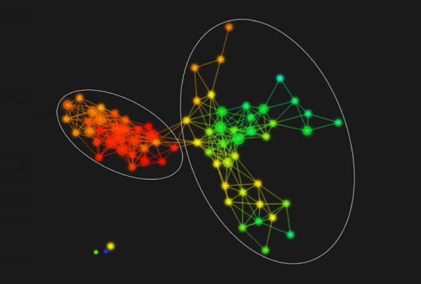
The researchers scanned 41 boys with fragile X syndrome using magnetic resonance imaging when the boys were roughly 3 and 5 years old. They then used an algorithm to find patterns in brain structure. They also measured the boys’ cognitive abilities, adaptive functioning and autism severity using standard scales.

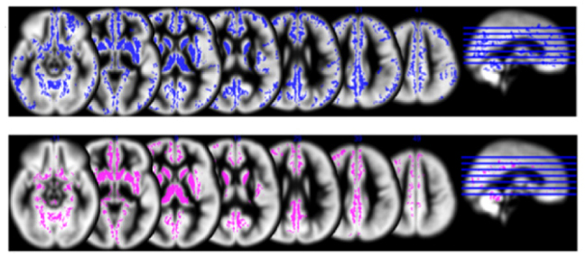
The algorithm identified two main structural types. Several brain regions are much larger in one type than in the other. These include the caudate, which is involved in learning; the thalamus, which relays sensory signals; and the cerebellum, which governs the coordination of movement. The brains of these children are also larger overall, particularly in the frontal and temporal lobes.
The results appeared 3 October in the Proceedings of the National Academy of Sciences.
Boys in the group with enlarged brains consistently score worse on all the behavioral and cognitive measures — meaning they are more severely affected — than do those in the group with smaller brains.
The affected regions are roughly the same as those found to be bigger in people with fragile X syndrome relative to typical individuals or people with autism alone. The enlargement in one subgroup may account for the apparent overall disparity, or this subgroup may have particularly large brains, says study investigator Jennifer Bruno, instructor in psychiatry and behavioral sciences at Stanford.
Stable subgroups:
To be sure the results reflect real subgroups, the researchers need to replicate their findings using a second algorithm, says Andrew Michael, assistant professor at Geisinger Health System’s Autism and Developmental Medicine Institute in Lewisburg, Pennsylvania, who was not involved in the study. “We want to make sure that [the finding] is not a result of the algorithm that they used, that it is truly biological,” he says.
The subgroups in the study appear to be stable: Boys with mild features at age 3 remained in that group at age 5. The researchers have rescanned the boys, who are now 12 and 13 years old, to see if those findings still hold. (The results are not published.) They also plan to match the early scans with later outcomes.
If a brain scan can reliably predict a child’s cognitive ability, it may help clinicians identify boys who are likely to benefit from early treatment, Michael says.
The team has launched a study of girls with fragile X syndrome. These girls typically display a wider range of features than boys with the syndrome, so scanning their brains may reveal additional subtypes of the condition.
References:
- Bruno J.L. et al. Proc. Natl. Acad. Sci. USA Epub ahead of print (2017) PubMed
Recommended reading

Expediting clinical trials for profound autism: Q&A with Matthew State

Too much or too little brain synchrony may underlie autism subtypes
Explore more from The Transmitter

Mitochondrial ‘landscape’ shifts across human brain

