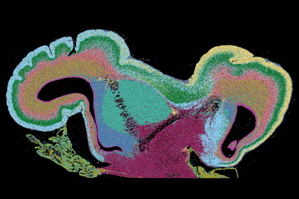
Brain atlas maps gene expression in three dimensions
Researchers have charted patterns of gene expression in a three-dimensional representation of the human brain. The results, published 20 September in Nature, show that different brain regions have distinct molecular and functional roles.
Researchers have charted patterns of gene expression in a three-dimensional (3D) representation of the human brain. The results, published 20 September in Nature, show that different brain regions have distinct molecular and functional roles1.
The map, called the Allen Human Brain Atlas, is based on two postmortem brains, one from a 24-year-old man and the other from a 39-year-old man, neither of whom had any signs of a neurological illness. The project aims to integrate information from as many as ten brains.
To create the map, the researchers first established a structure for the whole brain using magnetic resonance structural imaging. They then sliced the postmortem brains into thin sections and stained them with dyes and antibodies to identify populations of neurons and other brain cells.
The researchers isolated 900 discrete regions from these slices and cataloged the genetic messages that code for proteins in each slice.
Integrated into one picture, the data provide a 3D map of gene expression in the entire brain. Overall, the researchers found, gene expression varies significantly across the brain, with certain genes expressed only in discrete brain regions.
For example, genes related to dopamine signaling, which is involved in pleasure and reward, are expressed in a small subset of brain regions, including the striatum and the hypothalamus.
The researchers also looked at the expression of 740 genes that code for proteins in the postsynaptic density (PSD) — a neuronal region implicated in autism that receives signals across neuronal junctions, or synapses. About 30 percent of PSD genes are expressed only in a subset of brain regions, suggesting that variation in synaptic gene expression may underlie differences in brain regions.
Glia, support cells in the brain, express a number of PSD genes. Studies in the past few years have shown that glia may play a more significant role in brain signaling than was previously believed.
References:
1: Hawrylycz M.J. et al. Nature 489, 391-399 (2012) PubMed
Recommended reading

New organoid atlas unveils four neurodevelopmental signatures

Glutamate receptors, mRNA transcripts and SYNGAP1; and more

Among brain changes studied in autism, spotlight shifts to subcortex
Explore more from The Transmitter

Psychedelics research in rodents has a behavior problem
Can neuroscientists decode memories solely from a map of synaptic connections?
