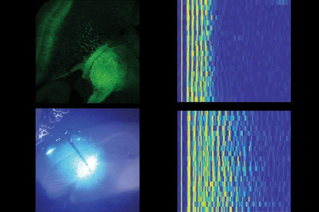
Autism mouse models show glitch in motor learning
Two well-known mouse models of autism show abnormal reactions to an eye-blinking test that relies on the cerebellum, a brain region that helps integrate sensory information and plan movements. The unpublished results were presented in a poster Monday at the 2012 Society for Neuroscience annual meeting in New Orleans.
A variety of methods have found cerebellar glitches in autism. For example, postmortem studies have found that individuals with the disorder have fewer Purkinje cells, which relay messages from the cerebellum to the outer layers of cortex, than controls do.
The new study suggests that the cerebellum does not work properly in mice lacking either CNTNAP2 or SHANK3, two leading autism candidate genes.
“We are bridging the gap between two areas in autism research — human brain defects and mouse models of autism,” notes lead investigator Samuel Wang, associate professor of molecular biology at Princeton University.
The findings are particularly exciting, he adds, given a study published earlier this year. That study showed that mice lacking the autism-related TSC1 gene only in Purkinje cells show social deficits and repetitive behaviors characteristic of the disorder.
“We believe that early-life disruption in the cerebellum can drive brain development off track in distant regions,” Wang says. “If that idea extends to the social brain, that would go a long way to explaining autism.”
In the new work, Wang’s team used a test called ‘eye-blink conditioning.’ The mice see a green light, which is followed by a puff of air on their eye. After hundreds of trials, they learn to close or partially close their eye after seeing the light, even when no puff comes.
People have the same reaction to the test. Interestingly, a study in 1994 found that individuals with autism show an abnormal response: They learn the association faster than controls do, but mistime it, blinking too early1.
“[The test] is the most well-studied paradigm that requires the cerebellum,” says Alexander Kloth, a graduate student in Wang’s lab who presented the work. The region is responsible for both how much the eye closes and the timing of the blink, he says.
SHANK3 mice don’t learn the blinking response as well as controls do, the study found. They also blink more quickly, with a later onset and earlier close, than controls do.
CNTNAP2 mice learn the response as well as controls do but, like people with autism, blink too early.
Kloth says it’s important to pay attention to these details of the response because they may correspond to circuit or gene expression differences in the cerebellum.
SHANK3, for example, is primarily expressed in a region of the cerebellum called the granule cell layer, which is involved in processing the light signal. CNTNAP2 is expressed in Purkinje cells. “We’re definitely not expecting the same result in every mouse model,” Kloth says.
For more reports from the 2012 Society for Neuroscience annual meeting, please click here.
References:
1: Sears L.L et al. J. Autism Dev. Disord. 24, 737-751 (1994) PubMed
Recommended reading
Explore more from The Transmitter


