Autism may alter how brain waves change with age
The strength and synchrony of brain waves appear to evolve differently in children with autism than in their neurotypical peers.
Brain activity patterns evolve differently in children with autism than in their neurotypical peers, according to a large study1.
Brain connectivity, a measure of the synchrony between brain areas, is known to be unusual in people with autism. But some studies have reported that brain connectivity is more synchronous than in controls, whereas others have reported that it is less so. The new study helps to reconcile these discrepant findings.
“It depends on the age at which one looks,” says lead investigator Margot Taylor, director of functional neuroimaging at the Hospital for Sick Children in Toronto, Canada. The study appeared in December in Annals of Neurology.
The researchers measured the strength, or power, and synchrony of brain waves. They found that in people with autism, the way in which these parameters evolve tends to be opposite to that seen in controls.
For example, in the cerebellum, a brain region linked to autism, brain waves of a certain frequency grow weaker until adolescence in children; in controls, they grow stronger in this time span.
The findings underscore the importance of taking age into consideration in brain imaging studies, says Tal Kenet, instructor in neurology at Harvard Medical School, who was not involved in the study. “You need to think carefully about whether you’re looking at something that has an age effect.”
Researchers should be particularly careful when conducting studies that involve adolescents, she says, because brain activity is more variable in adolescence than in early childhood.
Different development:
Taylor and her colleagues monitored brain activity in 61 children with autism and 73 typical children using magnetoencephalography. This method detects brain waves of various frequencies by measuring the magnetic fields produced when neurons fire. The children ranged in age from 6 to 16 years, allowing the researchers to look at age-related differences.
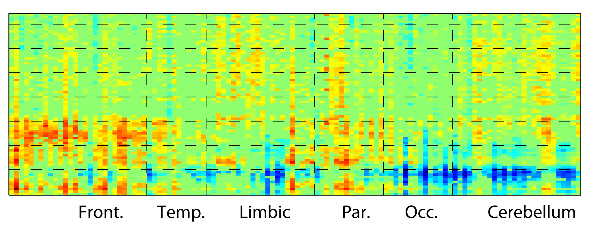
In typical children, medium-frequency brain waves known as alpha rhythms grow stronger with age until around adolescence, and become weaker after that. These patterns are seen primarily in three parts of the brain: the interior limbic regions, the occipital lobe and the cerebellum. In the frontal and parietal lobes, by contrast, low-frequency brain waves called delta rhythms and higher-frequency ones called beta rhythms become weaker with age until adolescence, and then grow stronger.
Brain waves in children with autism show opposite trends with age in these regions. Their alpha rhythms grow stronger at first and then become weaker, whereas their delta and beta rhythms grow weaker at first and then strengthen.
Out of sync:
The two groups also show age-related differences in the synchrony of brain activity within and between brain regions.
In controls, high-frequency gamma rhythms within the cerebellum, and between the cerebellum and the rest of the brain, become more synchronous with age. In people with autism, synchrony in these regions decreases.
Similarly, the synchrony of alpha rhythms throughout the brain decreases with age in controls, but increases in people with autism.
“Different frequencies in different parts of the brain are changing at a different rate in the two groups with age,” Taylor says.
The size of the study is impressive, says Elena Orekhova, researcher at the University of Gothenburg’s Gillberg Neuropsychiatry Centre in Sweden, who was not involved in the work.
But some of the differences seen between the two groups, particularly for the high-frequency brain waves, could stem from movement, which the study did not measure, she points out. Movement can affect rhythms, and children with autism are known to move more in scanners than typical children do.
It is also unclear how these differing trajectories relate to autism features. Taylor says her team plans to explore this question by including larger numbers of participants with a wider age range.
References:
- Vakorin V.A. et al. Ann. Neurol. Epub ahead of print (2016) PubMed
Recommended reading
Split gene therapy delivers promise in mice modeling Dravet syndrome
Changes in autism scores across childhood differ between girls and boys
PTEN problems underscore autism connection to excess brain fluid
Explore more from The Transmitter

U.S. human data repositories ‘under review’ for gender identity descriptors
