Martin Munz wanted to watch how the earliest cortical circuits form and become active in mammals. But he had a problem: Unlike the frog embryos he was accustomed to studying, which conveniently grow in transparent eggs, mammalian embryos develop inside a mother’s womb, making it difficult to spy on their growing neurons. Imaging the activity of these cells in living embryos, other researchers told him, was not feasible.
Munz got a more encouraging reply when he reached out to Botond Roska, director of the Institute of Molecular and Clinical Ophthalmology Basel in Switzerland. “He was like, ‘That sounds awesome; let’s do it,’” says Munz, now a postdoctoral researcher in Roska’s lab.
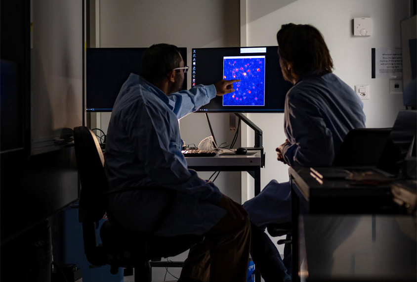
Munz teamed up with Arjun Bharioke, another postdoc in Roska’s lab, to develop this method. They describe the results of that seven-year-long effort today in Cell. Their novel method, which enabled them to stably visualize and record neuron activity in mouse embryos for five hours at a time, revealed previously unknown patterns of early cortical development.
The approach “opened up the way to study the early steps in the development of the cortex, which is fundamentally where people believe that these neurodevelopmental disorders would have an impact,” Roska says.
Mutations in two autism-linked genes prevent a temporary cortical circuit from forming at about embryonic day 14.5, the team found — a change that may shape later neuronal connections.
Three independent experts called the study a “tour de force,” including Xin Jin, assistant professor of neuroscience at the Scripps Research Institute in La Jolla, California, who added, “It’s the beginning of a very exciting chapter” for studying cortical development.
T
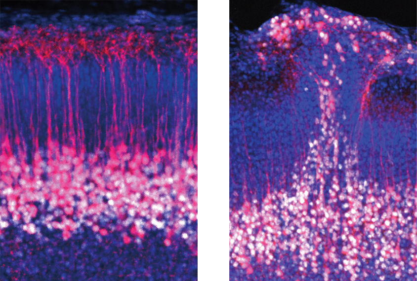
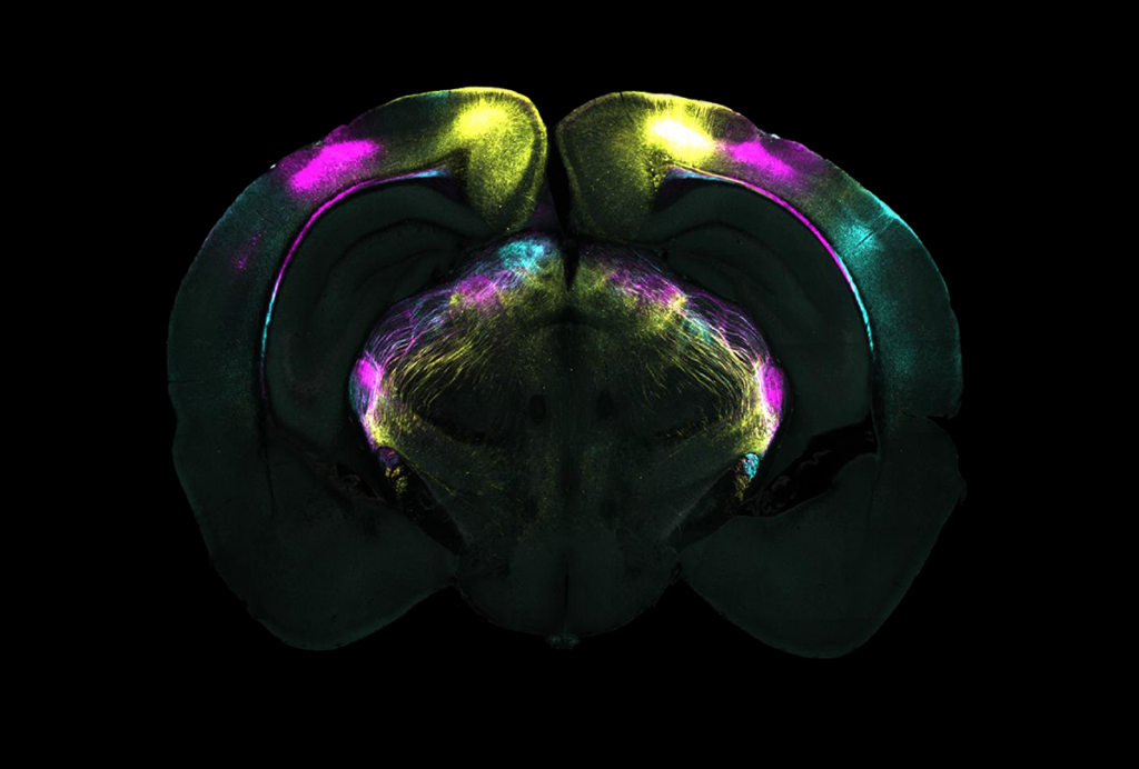
he mammalian cortex consists of six layers, each with characteristic connectivity. Cells in layers 2 and 3, for example, receive inputs from layer 4 and project down to layers 5 and 6, whereas layer 6 cells receive most of their inputs from upper layers and send signals out to the rest of the brain. Embryos form these layers from the bottom up, starting with layer 6 — the inner-most layer.Scientists have established how these early circuits develop mainly by studying the anatomy of brain tissue slices taken from embryos of varying ages. In recent years, teams have successfully measured neuronal activity in living embryos using two-photon imaging, but the methods can’t resolve fine-scale neuronal structures such as axons and dendrites.
Roska and his colleagues wanted to track when and how circuits form in a living animal, and they wanted to have the resolution to confirm that the circuits were functioning, Bharioke says.
But getting that high resolution is no small feat, Bharioke says. The team had to keep the live embryos warm and motionless for long experiments, so they placed the embryonic mice, still attached to umbilical cords, in an agar-filled container that sat partially inside the mother’s body. Two stabilizing arms held the container to limit the transfer of movements from the mother’s heartbeat and breath.
The team focused on the cells that ultimately form the cortex’s layer 5, which primarily contains pyramidal neurons that connect to one another and therefore do not have to rely on connections from other layers. These cells might offer one of the earliest glimpses into cortical circuit formation, the researchers hypothesized.
The cells spontaneously ramp up their activity between embryonic days 13.5 and 14.5 and then cluster into two sheets, forming a temporary circuit, the team found using two-photon calcium imaging and patch clamp recordings. The cells’ activity drops off for the next two days, and the temporary circuit disappears. At about day 16.5, the activity increases again, and by day 18.5, the cells have migrated to form the nascent layer 5, which persists at birth.
The location of activity changes within a cell over time, too — from the cell body toward the dendrites — as does the type of cells present, the team found.
“Before [these cells] become what we see in the adult, they seem to undergo a fairly complicated, convoluted developmental life,” says Oscar Marín, professor of neuroscience at King’s College London in the United Kingdom, who was not involved in the work. “It highlights that we know so little about in utero development,” he says.
Blocking the cells’ spontaneous activity prevents them from reorganizing into the two sheets by day 18.5, the team found. And embryos that carry a mutation in the genes CHD8 or GRIN2B, which significantly increases the likelihood of autism in people, do not form the typical clusters of cells at day 14.5 — suggesting that the autism-linked mutations alter the earliest stages of cortical circuit formation.
R
esearchers have known for decades that other groups of neurons form transient circuits during embryonic development, and the new work identifies another piece of that puzzle, says Zoltan Molnar, professor of developmental neuroscience at Oxford University in the U.K., who was not involved in the study.The imaging approach could be used to study other types of cells in other areas of cortex, and to investigate “how these transient circuits interact with one another,” Molnar says.
Roska and his colleagues plan to continue to investigate how these early circuits are established, and what problems arise when the temporary connections are eliminated. “What are the rules of transient circuits?” Roska says, and why are they made if they later disappear?
Figuring this out will ultimately require tracking how such circuit alterations affect behaviors, Molnar says, which is not something the researchers looked into in the study.
But the first priority is to characterize these early periods of typical development, he says, so that atypical development can be identified. “If we want to understand the origin of some of the developmental conditions, these are the stages which we have to study.”
