Autism gene wires social reward circuits in mouse brains
Mice with mutations in SHANK3, a leading autism candidate, may lack the neural wiring that would compel them to seek social contact.
Mutations in SHANK3, a leading autism candidate, cause mice to become solitary creatures rather than socialize with their peers. This is because SHANK3 wires the newborn mouse brain to seek social contact, suggests a new study1.
SHANK3 is known to strengthen the connections between neurons, known as synapses, in the mouse brain. The protein helps transmit signals that excite neurons. Mutations in SHANK3 weaken signaling among neurons, but how this translates to social behavior has been unclear.
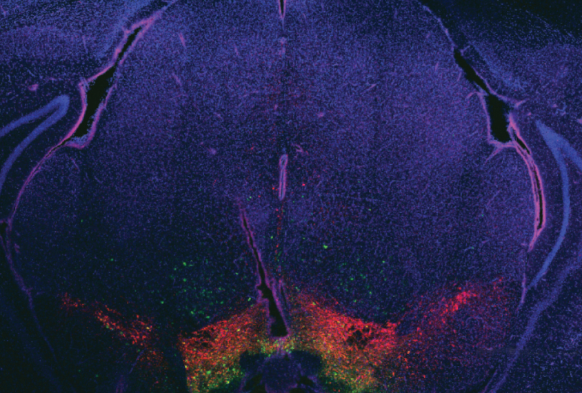
The study pins SHANK3 to a specific time and place in brain development. The researchers found that the protein connects neurons in the brain’s reward center, called the ventral tegmental area (VTA), during the first week of a mouse’s life. These neurons form a circuit that drives social behavior through adulthood, without further need for SHANK3. The research was published 6 June in Nature Neuroscience.
“This shows the combined roles of SHANK3 and the VTA in mediating social deficits,” says senior researcher Camilla Bellone, assistant professor of neuroscience at the University of Lausanne in Switzerland.
The findings lend support to the theory that, starting at an early age, children with autism are not motivated to seek others’ company. This in turn may limit their opportunity for social development.
Nearly 1 percent of people with autism carry mutations in SHANK3. The study points to the possibility of treating these people with molecules that target the receptor for glutamate, which mediates excitatory signals.
“This is the first study that implicates a special brain region and specific type of neurons in social impairments related to SHANK3 deficiency,” says Yong-Hui Jiang, associate professor of pediatrics at Duke University in Durham, North Carolina. Jiang was not involved in the study.
Early effects:
Bellone’s team dialed down the gene’s expression in the mouse VTA to levels seen in people with SHANK3 mutations. They used a synthetic RNA that silences the gene. This molecule blocks the gene’s expression in about half of neurons that produce dopamine in 6-day-old mice.
This creates a domino effect: Neurons in the VTA excite other cells — including those that produce dopamine, a chemical messenger involved in reward — much less than usual. As a result, the sense of reward that normally accompanies social interaction decreases.
The researchers waited until day 35, when the mice had completed adolescence and when synapse formation is normally complete. They then tested the neurons’ firing patterns using a technique called patch clamping. As expected, neurons that lack SHANK3 have less activity and less glutamate signaling in the VTA than control mice do, they found. The researchers attribute this finding to incomplete synapse formation.
The mutant mice spend more time grooming themselves, a common proxy for repetitive behavior associated with autism. The mice also spend only about five minutes on average with a cage-mate before scuttling off to an isolated area, compared with control mice which socialize for at least twice as long.
Receptor retrofit:
By contrast, silencing SHANK3 in early adolescence, around day 24, has no effect: Neuronal activity and social behaviors in the mice resemble those of the controls.
This suggests that, at least in the VTA, SHANK3 is most important in the first few days of life. This is also when the gene’s expression is at peak levels, Bellone says.
To explore whether the effects of SHANK3 silencing can be reversed, the researchers injected mice once daily, from day 6 through 18, with a molecule that activates the glutamate receptor — and can perhaps compensate for the low levels of glutamate.
The treatment works. It restores both signaling and social interest in the mutant mice, and these improvements persist through adulthood.
The researchers also used beams of light to stimulate dopamine neurons during the social interaction task, a technique called optogenetics. This treatment enhances social behavior, but only when mice encounter the light.
The study allowed the researchers to look at neural circuits specifically in the VTA. But this strategy might not mirror the full effect of SHANK3 mutations, Jiang says.
What’s more, the researchers saw a lack of social interest in the mice, which have SHANK3 levels similar to those seen in mice with one mutated copy of the gene. But mice typically do not show social deficits unless they have two mutated copies of SHANK3.
That finding is “extremely intriguing,” says Bellone. “Alterations in the VTA may represent an initial event that triggers downstream adaptations and leads to abnormal behaviors.”
Bellone plans to investigate how SHANK3 influences other neurons in the social reward circuit, including those that inhibit neuronal activity.
References:
- Bariselli S. et al. Nat. Neurosci. 19, 926-934 (2016) PubMed
Recommended reading

Too much or too little brain synchrony may underlie autism subtypes
Developmental delay patterns differ with diagnosis; and more
Split gene therapy delivers promise in mice modeling Dravet syndrome
Explore more from The Transmitter
During decision-making, brain shows multiple distinct subtypes of activity
Basic pain research ‘is not working’: Q&A with Steven Prescott and Stéphanie Ratté
