Feast your eyes on glowing glia and organoids; high-resolution, digital renditions of mouse brains; fluorescent beads passing through zebrafish guts and more.
Neuroscience—and science in general—is constantly evolving, so older articles may contain information or theories that have been reevaluated since their original publication date.
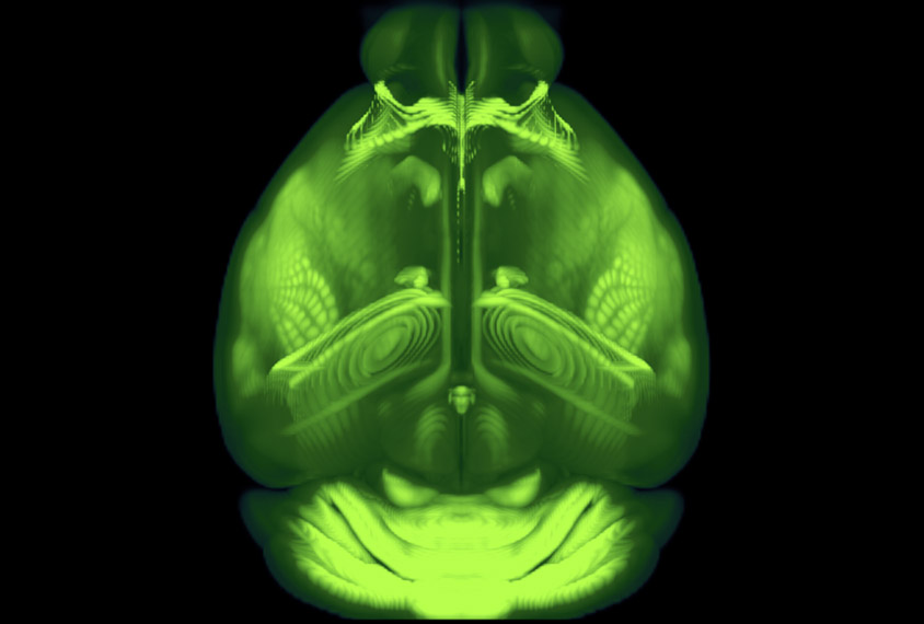
Precise pictures: This top-down view of the mouse brain is part of a new atlas that provides the most detailed 3D views to date. Researchers can use the reference to combine, analyze and share various types of mouse brain data.
Courtesy of the Allen Institute for Brain Science
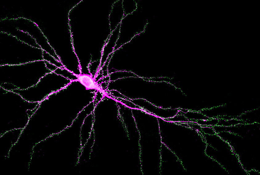
Bridging the gap: SYNGAP1 protein is located mostly at synapses, the junctions between neurons, (green) where it helps brain cells pass along chemical signals. People with a mutated copy of SYNGAP1 often have autism, as well as intellectual disability, epilepsy and an impaired gait.
Courtesy of Rick Huganir
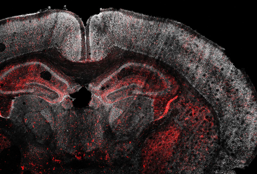
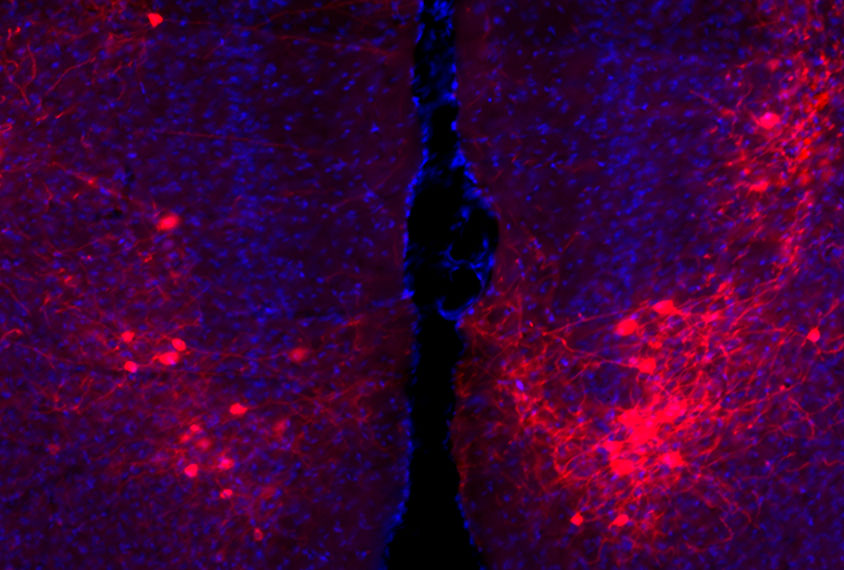
Cell by cell: A new technique called Perturb-seq makes it possible to screen for multiple autism-gene mutations (red), one by one, in living mice and to analyze their effects in individual cells.
Courtesy of Xin Jin / Harvard University
Drug targets: Fifteen different drugs commonly prescribed to people with autism created unique behavioral signatures in mice.
Courtesy of Alex Wiltschko / Harvard University
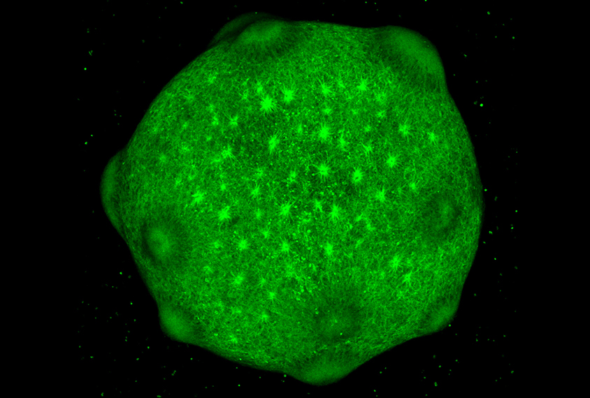
Electric organoid: Neurons derived from people with 22q11.2 syndrome are hyperexcitable and show calcium-signaling deficits.