Touch sensors detect subtle environmental vibrations, send information to auditory midbrain
Pacinian corpuscles sense high-frequency vibrations from meters away and send the information to a different circuit than other touch signals, according to a pair of new studies.
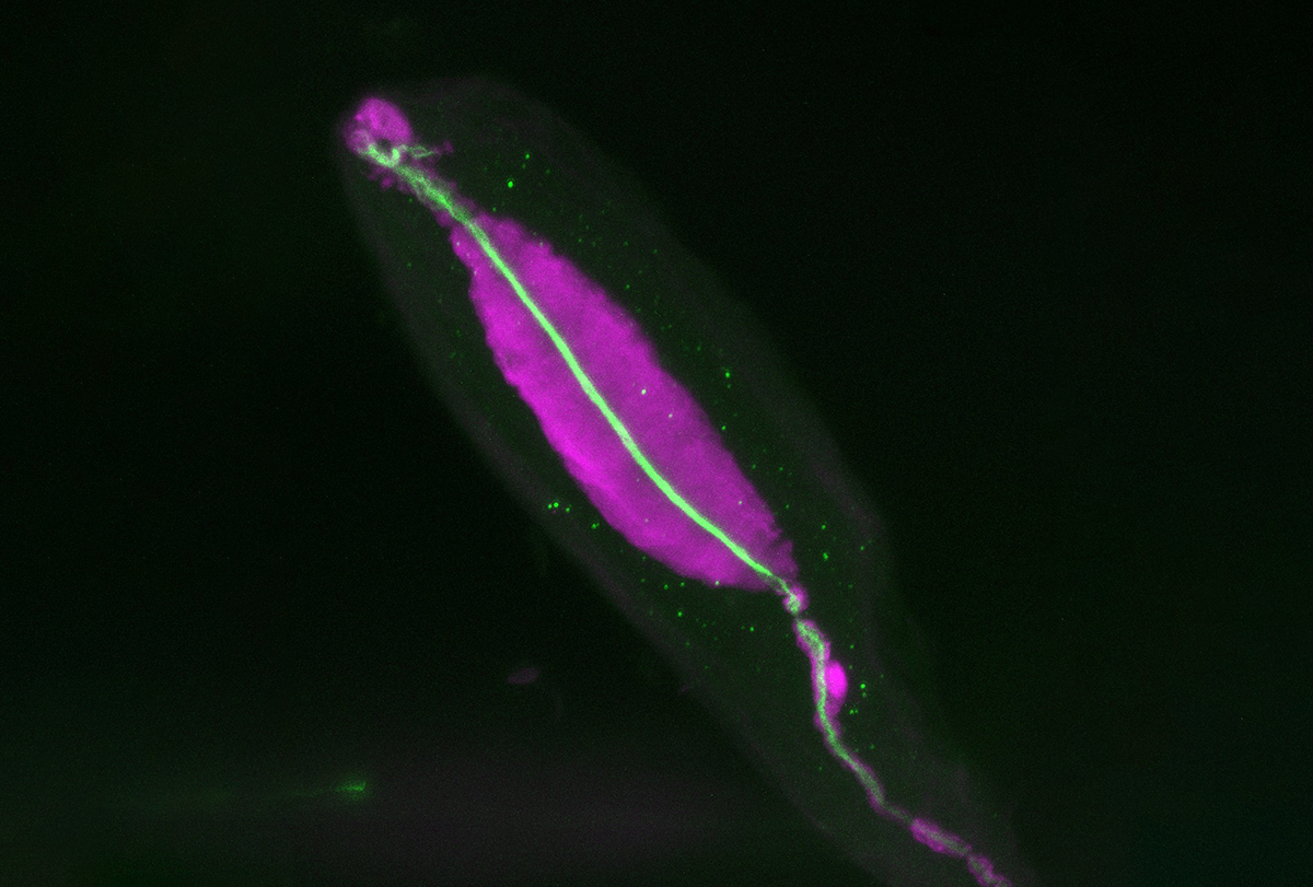
Spinal tap: Recording from dorsal root ganglion neurons in freely behaving mice revealed Pacinian corpuscles, shown here, are more sensitive than previously thought.
Courtesy of the Ginty Lab
Neurophysiologist Josef Turecek learned a valuable lesson last year: If you need to carry something strange across a hospital campus, put a tape measure on your belt.
Turecek, a postdoctoral fellow in David Ginty’s lab at Harvard Medical School, was studying how somatosensory neurons respond to various forms of touch. Turecek had spent about six months developing a way to record from the neurons’ cell bodies in the dorsal root ganglion, a ball of cells dangling off the spinal cord, in freely behaving mice. Now he was testing the sensitivity of the neurons’ axons in the skin, where some of them use a sensor called a Pacinian corpuscle to detect vibration.
He placed mice on different surfaces—sandpaper, foam, cardboard—and gently dragged his finger across the surface, without touching the mouse. The Pacinian neurons fired in a series of staccato pops, indicating they detected surface vibrations. This was intriguing, Turecek says, because the classic literature had only demonstrated that the neurons detect vibrations applied directly to the skin. So Turecek decided to test a surface that mice encounter in the wild: a tree branch. Which meant he needed to carry an 8-and-a-half-foot-long stick across the Harvard Medical School campus.
He looked like “the Grim Reaper,” he says, but because he had a tape measure on his belt, “everyone was fine with it.”
Back in the lab, Turecek nestled the branch into the small, dark recording room. He placed the mouse on one end of the branch and gently tapped the other end. The Pacinian neurons fired.
The neurons also fired right after the mouse’s foot made contact with the ground, in the absence of surface vibrations. This may be due to the location of Pacianan corpuscles, says Ginty, professor of neurobiology: In humans and monkeys, Pacinian corpuscles are located right under the skin and in connective tissues; in mice, they reside in the membrane wrapped around ankle and wrist bones. Vibrations from making contact with the ground may resonate through the skeleton and activate the Pacinian corpuscles, according to a paper Ginty and Turecek published 7 August in Neuron.
“The neurons turned out to be even more sensitive and respond to even more external stimuli than we had imagined,” Ginty says. “We never would have known that had the animal not been standing on its feet and walking around while we were recording.”
Good vibrations: Pacinian neurons fire in response to surface vibrations travelling through sandpaper, cardboard, foam and a tree branch.
Turecek and Ginty, Neuron 2024
Recording from neurons in the dorsal root ganglion was a “technical breakthrough,” Ginty says. In the classic literature from the 1960s and 1970s, researchers peeled back a section of skin to expose the sensory nerves and their various end organs. Then they stimulated the skin with different types of mechanical force and measured what each end organ responded to. More recent studies record from sensory nerves while stimulating the foot of anesthetized mice. But there has been a “gap” between information gleaned from those methods and “what that actually means in terms of behavior and natural stimuli,” Turecek says.
It’s difficult to record from neurons in the spinal cord, because things move around a lot, Turecek says: “The body is meant to be twisting and turning.” To create a stable area, Turecek fused three vertebrae together in the spine, in addition to other feats of engineering problem-solving. “People always asked me, ‘What was the thing that allowed you to do it?’ And it’s not really one thing; it’s 100 things have to go perfectly right.’”
This approach is “very, very interesting,” says Slav Bagriantsev, associate professor of cellular and molecular physiology at Yale University, who was not involved in the work. “By doing this kind of experiment, you can ask questions that you cannot ask through other skin-nerve preps.”
T
he brain may process some Pacinian-encoded vibration information differently than other forms of touch, according to a preprint the Ginty lab posted on bioRxiv in March. In the canonical pathway, information flows from dorsal root ganglion sensory neurons to the dorsal column nucleus in the brainstem, which then sends projections to the thalamus and then the somatosensory cortex.
But many of the neurons that respond to high-frequency vibrations project to the inferior colliculus—an area of the midbrain involved in auditory processing—and not the thalamus, Turecek observed while recording from dorsal column nucleus cells in an anesthetized mouse.
“Right away, we just knew that this was something that was intrinsically interesting and unexpected,” says study investigator Erica Huey, a graduate student in Ginty’s lab.
To expand on Turecek’s observation, Huey recorded from neurons in the inferior colliculus and thalamus while stimulating the foot of an anesthetized mouse with vibrations ranging from 10 to 900 hertz. Most neurons in the thalamus had the strongest responses to vibrations less than 200 hertz, whereas neurons in the inferior colliculus preferred vibrations greater than 200 hertz, Huey found.
This division may be due to the fact that the two pathways receive input from different vibration-sensing end organs, Huey discovered in a series of genetic knockout experiments. In mice without Pacinian corpuscles, the inferior colliculus barely responded to any vibration frequency, but the thalamus still did. Knocking out Meissner corpuscles, another vibration sensor, also did not affect vibration sensitivity in the thalamus, which indicates the area receives low-frequency vibration information from multiple types of mechanosensory neurons.
In the inferior colliculus, about 75 percent of neurons fired in response to both vibration and sound, and most of those neurons had a stronger response when both stimuli occurred at the same time. When Huey inhibited the inferior colliculus with muscimol—a GABA receptor agonist—awake, head-fixed mice no longer preferred a stationary platform over a vibrating one, but they still avoided unpleasant textures and temperatures.
This makes sense, because both “sound and touch are vibrations,” says Manuel Sánchez Malmierca, professor of histology at Universidad de Salamanca, who was not involved in either study, so it follows that “neurons which are sensitive to sound frequencies are also sensitive to vibrations.” This double encoding may provide redundancy to ensure mice avoid threats, Turecek says: If a mouse both hears and feels an approaching predator, they will pay more attention to it.
The researchers may have missed some response to high-frequency vibrations in the canonical pathway because of the thresholds they selected for what constitutes a response, says Kuo-Sheng Lee, assistant research fellow at Academia Sinica, who was not involved in the work. Lee and his colleagues mapped out the vibration receptive fields in each brain area along the canonical pathway and found that some cells do tune in to high frequencies, according to a preprint posted on bioRxiv last September.
But regardless, the conclusions of the preprint stand, Lee says: The inferior colliculus pathway may drive detecting and avoiding vibration in the environment—whether it comes in the form of sound waves or mechanical vibrations—whereas the canonical pathway processes vibrations that are relevant to discriminatory touch.
In future work, Ginty and Turecek say they hope to dig into how the brain processes the high-frequency vibration information after it leaves the inferior colliculus.
“The historical view would have been, ‘Cortex is involved in the perception and behavior in response to vibrations,’” says Jeffrey Yau, associate professor of neuroscience at Baylor College of Medicine, who wasn’t involved in either study. “And so in the mouse, I think this is actually raising an interesting question of, if that high-frequency information isn’t necessarily going through thalamus and getting to cortex, then what is cortex doing with respect to these signals?”
Other ongoing projects include exploring how vibration is represented in the auditory pathway of deaf animals, applying the dorsal root ganglion recording technique to other sensory end organs, and comparing how the structure of different end organs relates to their function—both within and between end organ types. Pacinian corpuscles, for instance, come in a range of sizes: Some look like a carrot, whereas others resemble a chili pepper or potato, Turecek says. Differences in shape tweak the specific range of vibrations it responds to. “Every Pacinian is a little different.”