Null and Noteworthy, relaunched: Probing a schizophrenia biomarker
This edition of Null and Noteworthy—the first for The Transmitter—highlights new findings about the auditory steady-state response in people with schizophrenia that, all within one study, somehow packed in a null result and a failed replication.
Welcome to Null and Noteworthy, a monthly newsletter about neuroscience research that fails to support a hypothesis or replicates a previously proposed one. Though these studies are typically less splashy than those that many journals publish, they often bring more nuance and complexity to the field by exposing cracks in or solidifying previous findings.
We originally launched this newsletter in 2021 for Spectrum, and now we’ve revamped it for TheTransmitter. The study highlighted below somehow packed into a single paper a null finding, several replications and a failed replication—a microcosm of the scientific and publishing processes in action.
Have a notable null result or replication we should cover, or other suggestions for this newsletter? Send your tips to [email protected].
Sound off:
A repeating sound prompts oscillating waves of brain activity known as the auditory steady-state response (ASSR)—most strongly for sounds that occur 40 times per second. The power of this response is dulled in people with schizophrenia, according to a seminal 1999 paper and numerous replications over the past two decades, suggesting it might serve as a biomarker for the condition.
Blocking NMDA receptor function with drugs such as ketamine blunts the response in laboratory animals, other studies show, suggesting that the receptors contribute to some of schizophrenia’s traits.
Similar alterations occurred on two measures of the 40-hertz ASSR in a group of 31 neurotypical people who received ketamine compared with when they got a placebo, according to a new study that replicates prior results: The participants’ ASSR showed less phase variability and reduced power with ketamine. The same shifts were seen among 51 people with schizophrenia or schizoaffective disorder included in the study—the first to directly compare schizophrenia and ketamine, removing possible confounding effects from experimental design in previous comparisons across studies.
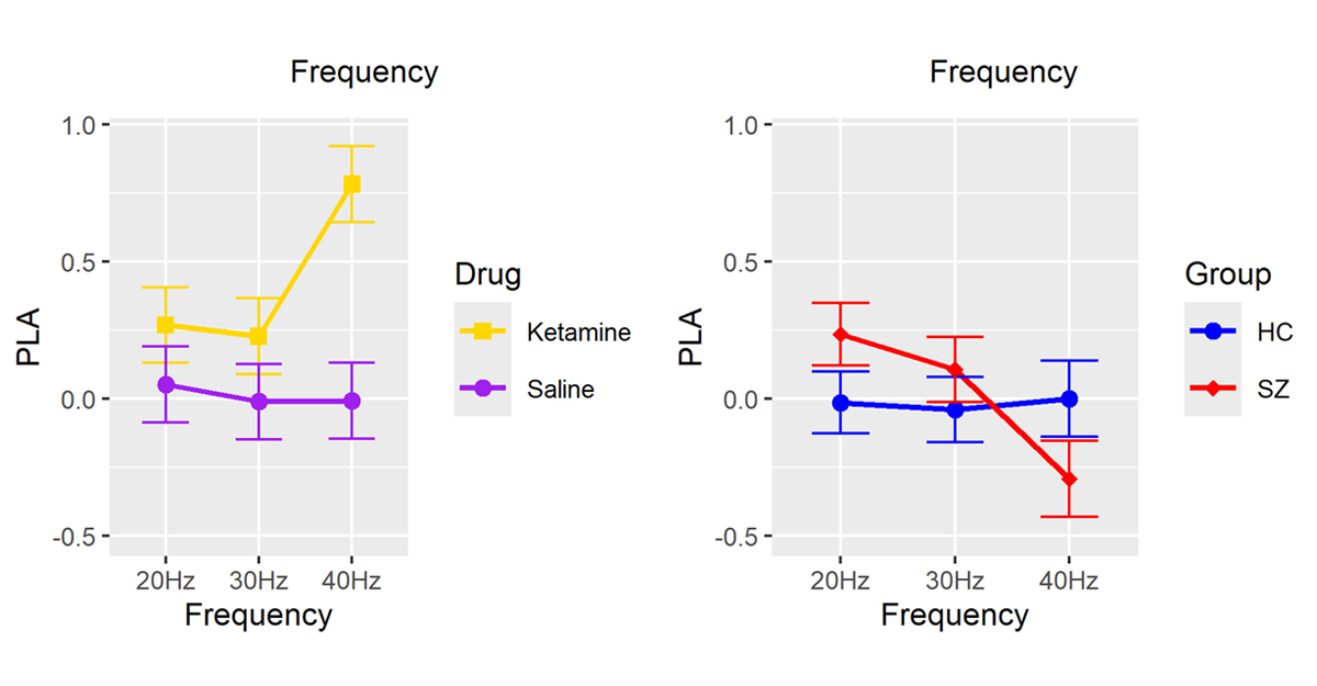
Signal split: People receiving ketamine infusions (left, yellow) respond more quickly to a 40-hertz sound than does a control group (left, purple). The same response is slowed in people with schizophrenia (right, red) compared with controls (right, blue).
But on a third measure of the time it takes for the brain to respond to the sound, called the phase-locking angle, the responses of participants with schizophrenia were not significantly different from those of an independent group of 52 neurotypical controls—contrary to previous studies that showed a greater delay in people with schizophrenia. The new work was published in January in Biological Psychiatry: Cognitive Neuroscience and Neuroimaging.
What’s more, the response was sped up in people who received ketamine compared with controls, suggesting NMDA receptors can’t fully explain alterations to the 40-hertz response in people with schizophrenia, says study investigator Brian Roach, a data scientist at Carrum Health. (At the time of the study, Roach was a research scientist at the nonprofit Northern California Institute for Research and Education.)
It’s possible that the change in phase-locking angle in schizophrenia is small and requires a large sample size to detect, says Molly Erickson, assistant professor of psychiatry and behavioral neuroscience at the University of Chicago. In a 2022 study, she and her colleagues found a significant delay in 78 people with schizophrenia compared with 80 controls. But that delay also appeared in Roach’s 2019 study in just 28 people with schizophrenia and 25 controls, suggesting sample size is not the culprit, Roach says.
And Roach’s team has unpublished data showing further phase advancement in people receiving ketamine, he says.
Rather, the culprit could be individual differences among people with schizophrenia, Roach says, potentially undermining the 40-hertz ASSR’s usefulness as a biomarker. “If what you’re looking for from a biomarker is that it consistently shows you an abnormality, that’s not what we’re seeing so far.”
Roach’s co-investigator is currently examining the response in larger cohorts, including one of children at high risk of schizophrenia, Roach says. “If it is an issue of power and effect size, that will certainly play out in those larger datasets.”
Et al.:
A lack of microglia doesn’t disrupt a host of functions thought to rely on the cells, including synapse maturation, seizure susceptibility and neuronal gene expression, according to a study in mice. The Transmitter previously covered emerging questions around the true role of microglia, particularly in early development. Nature Neuroscience
Carriers of apolipoprotein E-4—a genetic risk factor for Alzheimer’s disease—encode memories just as effectively as people with other variants of apolipoprotein E do, according to study that used a pre-registered movie-watching method. Brain and Neuroscience Advances
Facial expression and facial redness have no effect on an observer’s brain response in the occipital area that occurs shortly after the brain begins processing visual information. eNeuro
Editors of a series on replications at Nature Communications rejected a paper replicating previous findings on episodic memory because of questionable statistical concerns, according to Stuart Buck, executive director of the Good Science Project, a nonprofit think tank focused on U.S. science funding; the paper was ultimately published in iScience. The Good Science Project
Performance on tests of scene recognition, spatial memory and navigational abilities is not associated with resting-state functional connectivity between brain regions responsible for processing visual information from the environment. eNeuro