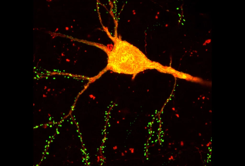
Hunched over a microscope more than a century ago, Santiago Ramón y Cajal discovered that distinct types of neurons favor different brain regions. Looking at tissue from a pigeon’s cerebellum, he drew Purkinje cells, their dendrites outspread and twisted like a ravaged oak. And drawing from another sample—the first cortical layer of a newborn rabbit’s brain—he traced the tentacled nerve cells that would later bear his name.
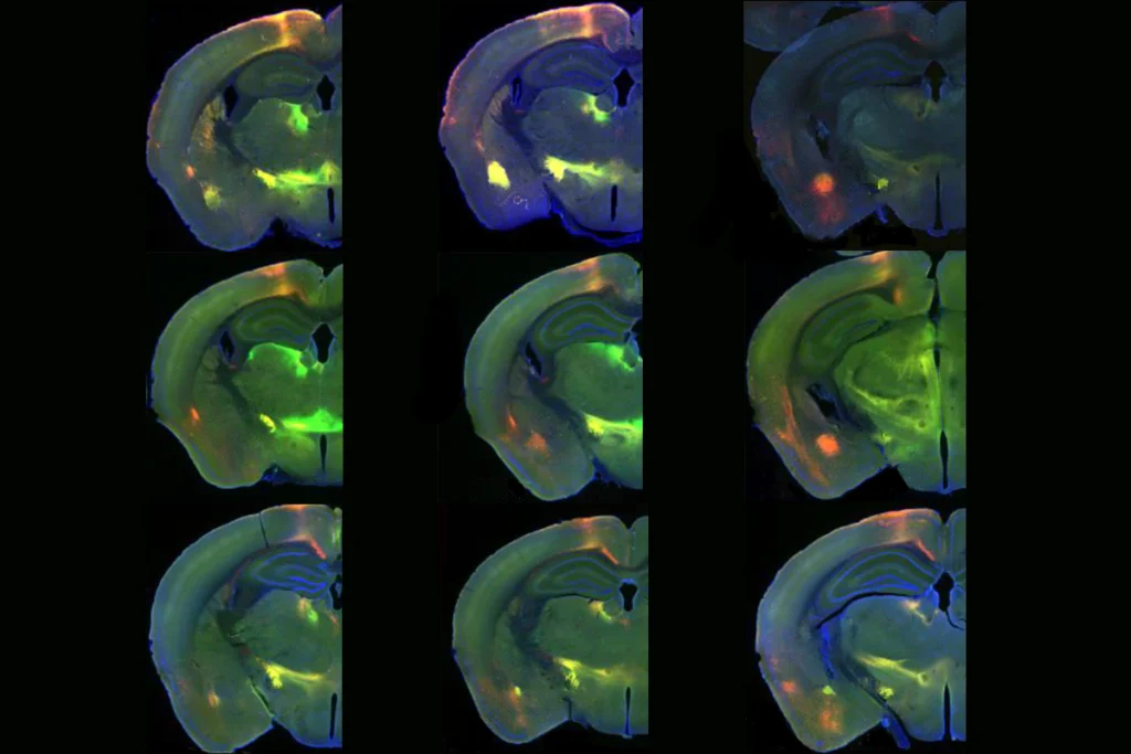
But the brain’s cellular organization is even more ordered than Ramón y Cajal could have imagined, a new study suggests. Different functional networks—measured using functional MRI—involve distinct blends of cell types, identified from their transcriptional profiles. And a machine-learning tool trained on cell distributions in postmortem tissue can identify functional networks based on these cellular “fingerprints,” the researchers found.
The findings could address the gulf between neuroimaging and cell-based research, says the study’s principal investigator, Avram Holmes, associate professor of psychiatry at Rutgers University. “In-vivo imaging studies are almost never linked back to the underlying biological cascades that give rise to the phenotypes,” he says. But the new approach “lets you jump between fields of study—that was very difficult to do in the past.”
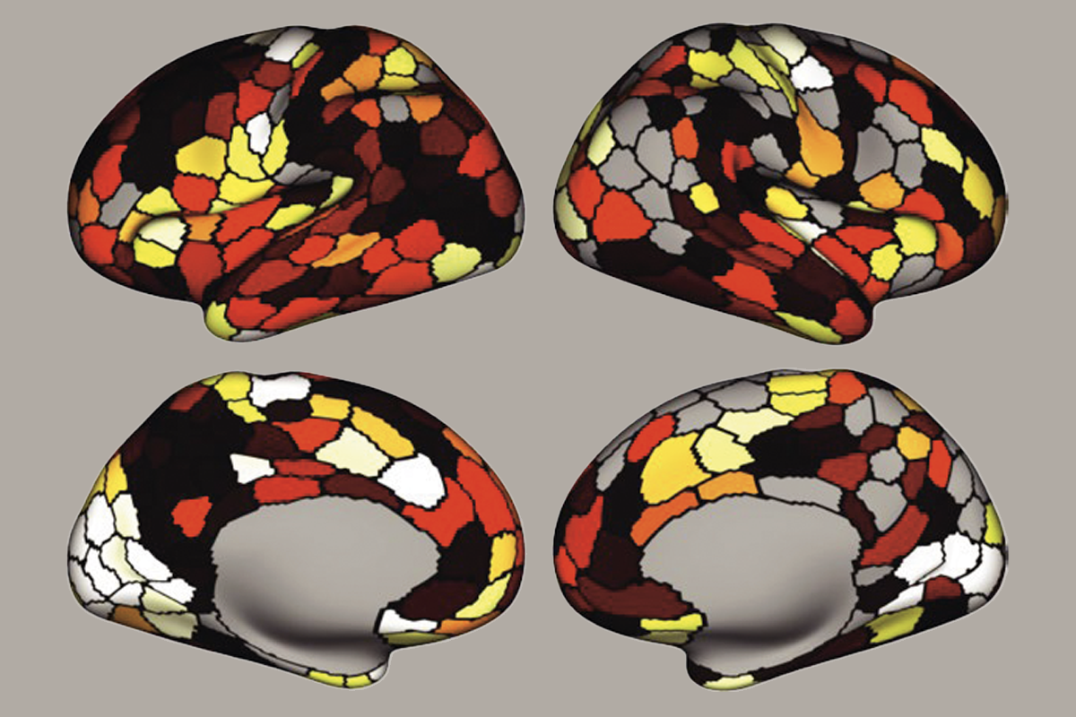
Using bulk gene-expression data from postmortem human brain tissue—obtained from the Allen Human Brain Atlas—Holmes and his colleagues classified 24 different types of cells. They then mapped the cells’ spatial distribution to two features of large-scale brain organization derived from a popular fMRI atlas: networks, and those networks’ position in the cortical gradient, which is based on location, style of information processing and connectivity pattern. Unimodal sensorimotor networks—those that perceive stimuli and act on them—anchor one end of the gradient, and the other end is occupied by transmodal systems, such as the default mode network, that integrate multiple information streams across the cortex. The remaining networks are parked between these two extremes.
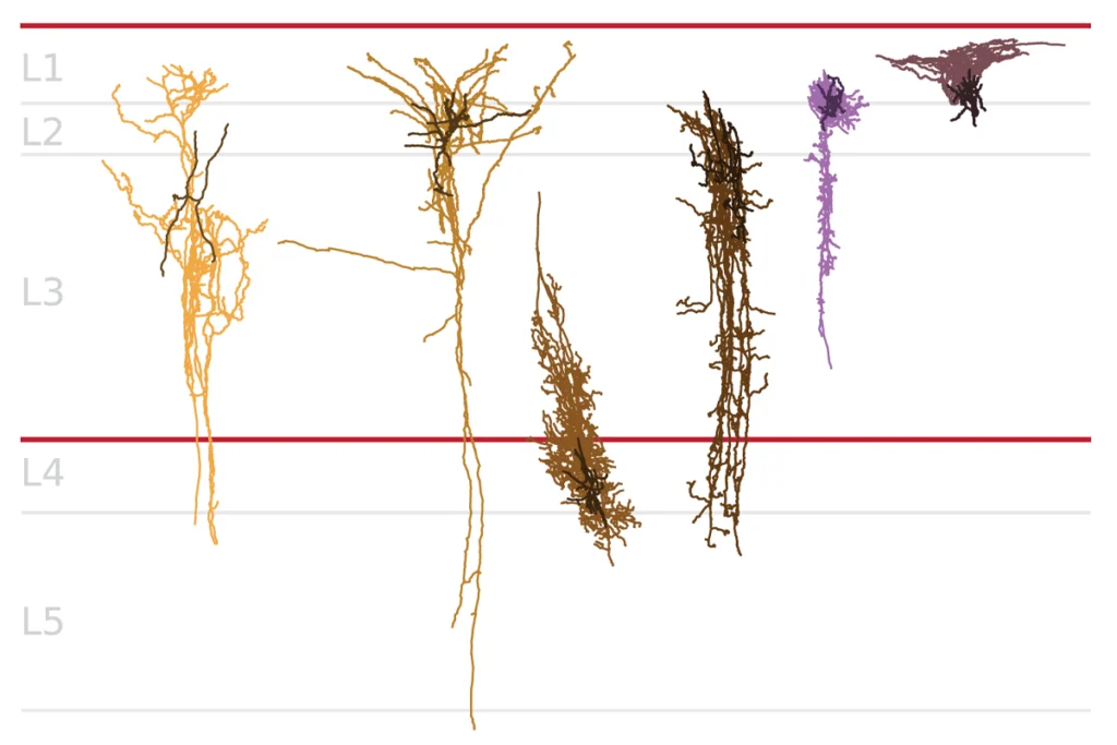
Different cell types align with different parts of this gradient, the researchers found. For instance, somatostatin-expressing inhibitory interneurons are more abundant among transmodal networks, whereas layer 4 intratelencephalic excitatory neurons are more common at the unimodal end.
And the ventral attention network’s cellular configuration—which sits in the middle of the gradient—looks like a combination of both extremes. Its composition is so distinctive that a computer model trained on cell type composition from postmortem brain tissue can identify the network certain cells belong to, the researchers found. The work was published in November in Nature Neuroscience.
T
hat particular mix of cells might contribute to the network’s role in neurodevelopment: During puberty, networks mature along the cortical gradient, with complex cognitive skills developing last. That process is associated with the level of connectivity within the ventral attention network, according to a study published by Holmes’ group last year. Probing further into its cellular signature might reveal how the ventral attention network prompts cortical maturation, Holmes says.The findings could also help scientists understand the spatial patterning of neurological conditions, says Konrad Wagstyl, senior lecturer in biomedical computing at King’s College London, who was not involved in the work. Brain scans of people with neurological conditions sometimes show alterations that map onto the cortical gradient. Understanding spatial organization at a cellular level could help explain that pattern, he says.
Whereas Holmes’ team selected networks from a single brain atlas, future studies are expected to uncover how well cellular organization aligns with other maps, which sometimes categorize the same networks slightly differently, says Lucina Uddin, professor of psychiatry and biobehavioral sciences at the University of California, Los Angeles, who was not involved in the work. “It will be interesting to see how the findings replicate across multiple different functional MRI-based atlases.”
And cataloging cell types isn’t perfect either, Holmes admits. Because methods to define both cellular identities and networks are in their infancy, his team has likely underestimated the relationship between these levels of brain organization, he says. “As other techniques are refined, I’m anticipating we’ll get a much cleaner picture of how cellular architecture relates to functional properties.”
Holmes and his team have posted the code for their analysis online so that other groups can repeat the pipeline using their preferred atlas. “Our hope is that people apply it to their modeling approach, so you end up with spatially informed models of how the cellular properties of the cortex affect its functions,” Holmes says.