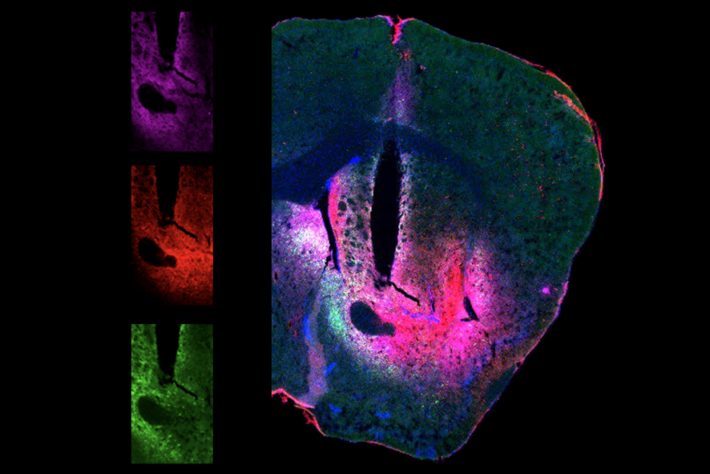
Several years ago, Tony Kelly and his colleagues thought they were onto something major: They had equipped their mouse model of epilepsy with a calcium sensor that fluoresces when neurons fire, and imaging revealed striking patterns of light that crawled across the hippocampus as the animals experienced seizures.
“We were like, ‘This is it,’” says Kelly, a researcher at the University of Bonn.
But their excitement quickly shifted to confusion and disappointment when they saw the same wave patterns in wildtype animals. And Kelly and his colleagues soon found out they were not alone in being fooled by the activity, which they termed “micro-waves.” In fact, several other labs who use calcium imaging in the hippocampus had observed these same waves for more than a decade.
Lab by lab, researchers had come to the conclusion that the waves were spurious and had changed their protocols accordingly, Kelly says. To reach more people, he and his colleagues decided to publish their artifactual findings as a cautionary tale in eLife in April. They say the calcium imaging technique is still useful—and offer tips on how to keep recordings artifact-free.
Documenting this hippocampal micro-wave artifact is important for the field—particularly as calcium imaging becomes more affordable and more labs try their hand at it, says Mark Sheffield, assistant professor of neurobiology at the University of Chicago, who came across the same artifact about 10 years ago as a postdoctoral researcher.
“Otherwise, we’re all figuring out all the negative results independently, [using] resources, money [and the] lives of our animals that we work with,” he says. “It’s just a waste.”
C
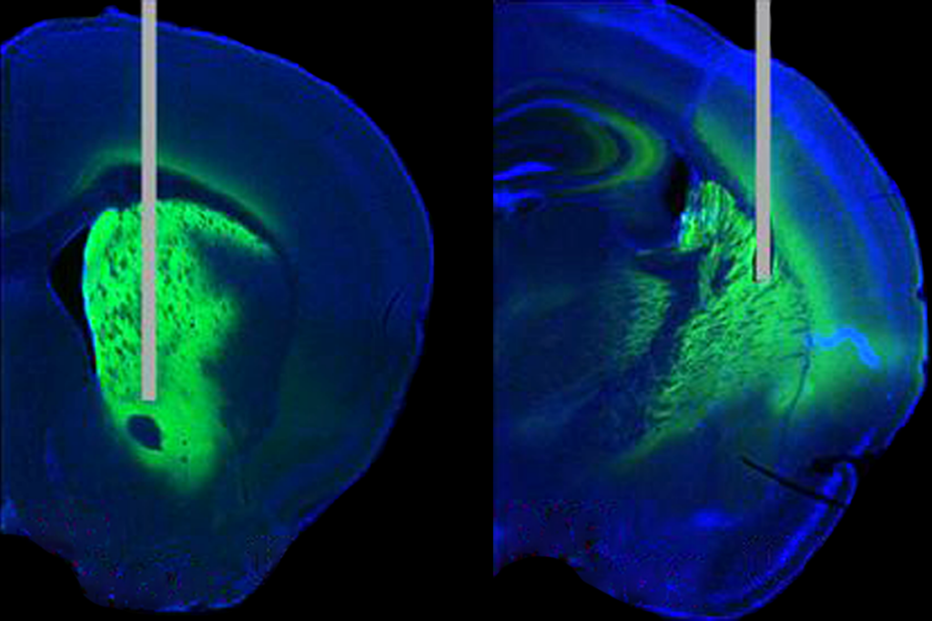
alcium imaging relies on a genetically encoded calcium indicator—a protein that fluoresces when it binds to calcium ions, which rise inside a neuron when it fires. Researchers can use fluorescence microscopy techniques to track the cells that light up.Many labs, including Kelly’s, typically deliver the indicator gene into an animal’s cells using a virus injected into the brain. A promoter—often one that targets the synapsin protein, which is specific to neurons—drives the indicator’s expression in only the desired cell types.
But in certain circumstances, the calcium indicator can create problems, Kelly and his colleagues found when they tested the protocol across multiple labs. Paired with the synapsin promoter, many indicators, including GCaMP6m, GCaMP6s, GCaMP7f and R-CaMP1.07, gave rise to the aberrant waves in the hippocampus—but not in the cortex. Those waves typically showed up around four weeks after viral injection and happened more frequently when cells were injected with the highest volumes of virus in a typical protocol, the team found.
The waves did not occur, however, when the team used the promoter CaMKII, which targets excitatory but not inhibitory neurons. They also did not show up in transgenic mice that were genetically modified to express the calcium indicators, rather than having the indicators inserted virally.
It is still unclear why these artifacts occur, Kelly says. He and his colleagues did not seek to explain the phenomenon in the new paper, only to characterize them, he says.
One long-standing hypothesis is that the waves have little to do with the indicator or the promoter; instead, they are a reaction to the tissue being damaged, says Daniel Dombeck, professor of neurobiology at Northwestern University, who was not involved in the work. He says he came across the waves when his lab was imaging the hippocampus more than a decade ago, typically with researchers who were new to calcium imaging.
“Once people get very good at the surgeries and the injections, it’s pretty rare to see these things,” Dombeck says. Experienced labs develop protocols for dealing with the artifacts when they do arise, including scrapping the experiment and moving on to a different animal, he adds. “If we see the waves, we know that something went wrong.”
Some researchers even have a name for the artifacts, says Loren Looger, professor of neurosciences at the University of California, San Diego, who was not involved in the new work. “We and others have been talking for 15 years about cells getting ‘GCaMPed out,’” he says.
And the problem may also affect the cortex—contradicting Kelly’s findings. Unusual cortical neuron activity has been reported in both transgenic and virally infected mice expressing calcium indicators long-term, two different teams have reported.
“Lots of things mess up cells after long-term expression,” from GCaMP to optogenetic reagents and other sensors, says Looger, who led one of those studies. Although he says he appreciates more research into the unusual activity, “it’s not at all surprising that things get messed up when we express something in our system.”
T
he artifacts should not prevent researchers from using calcium imaging, Kelly says. “In no way would I like to cast any aspersions on anyone who’s used this technique, because I think it’s an amazing technique. It’s really, really useful. And I think most people probably do use it properly.”In the new paper, Kelly and his colleagues recommend that researchers who perform calcium imaging in the hippocampus take steps to avoid these artifacts, such as using a promoter other than synapsin, or keeping the volume of the virus low.
He also does not blame other researchers for not publishing about the artifacts in the past. “Publishing negative data takes time. And it’s not so easy to do,” he says.
But as calcium imaging has become more affordable, the number of researchers interested in using the technique has blossomed—meaning an increase in the number of people coming across these micro-waves, Sheffield says. He adds that he has never seen mention of them in published papers, but he has seen them on posters at the annual Society for Neuroscience meeting.
Dombeck says that he and his colleagues have made a point of spreading the word about the artifacts over the past decade. But publishing a comprehensive look at the artifacts and the protocols that lead to them, as Kelly and his colleagues have done, will help educate people more widely, he says. “It’s a service to the community. It’s a great thing.”


