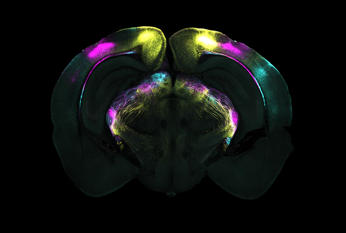
Circuit training: Neuronal projections (shown in pink, yellow and blue) from the visual cortex to the vLGN help mice learn to suppress instinctive fear responses.
Subthalamic plasticity helps mice squelch innate fear responses
When the animals learn that a perceived threat is not dangerous, long-term activity changes in a part of the subthalamus suppress their instinctive fears.
Mice have poor vision, but they still instinctively run or freeze if they see a potential threat, such as the shadow of a looming predator. They can override these instincts, though, if they learn over time that a perceived threat is harmless.
The ventrolateral geniculate nucleus (vLGN) of the thalamus, which receives visual information from the retina, helps to guide these reflexive behaviors. But the vLGN does more than meets the eye, or retina, according to a study published today in Science. It also stores vision-dependent fear memories and undergoes experience-dependent plasticity to suppress fear behaviors when mice learn that a perceived visual threat is not actually dangerous.
“It was known that [the vLGN] received strong input from the eye, from the retina, but it was quite mysterious what it actually does,” says study investigator Sonja Hofer, professor of neuroscience at the Sainsbury Wellcome Centre for Neural Circuits and Behaviour.
Fear learning and memory is typically associated with the hippocampus, amygdala, cerebellum and prefrontal cortex. Finding it in the vLGN “is, in my opinion, quite surprising and very interesting,” says Newton Sabino Canteras, professor of anatomy at the University of São Paulo’s Institute of Biomedical Sciences, who was not involved in the research. “And this opens to other situations where we have subthalamic regions modulating this kind of response.”
I
n a commonly used fear-memory paradigm, mice escape to a sheltered area in response to an expanding circular shadow projected into their enclosure to simulate an approaching predator.
Courage creation: Mice flee in response to looming predator shadows but over time learn to suppress this instinctive behavior.
Courtesy of the Sainsbury Wellcome Centre
After the mice escape, they show a sharp increase in the activity of vLGN neurons that project to the superior colliculus, a structure that helps orient motor responses, Hofer and her colleagues discovered in 2021. But after seeing the stimulus multiple times, the mice are less likely to escape, and the activity of the vLGN neurons drops. Suppressing those neurons via optogenetics caused the animals to flee, whereas activating the neurons blunted the animals’ fear reactions, the study also showed. The results suggested that experience modulates activity in the vLGN, so the researchers began trying to work out the specifics of this pathway.
In the new work, the team used the same behavioral assay, but after exposing the mice to the shadow, they installed a barrier to prevent animals’ escape and continued exposing them to the looming shadow. Once the researchers removed the barrier, the mice rarely fled to the shelter, suggesting they had learned the shadow was harmless and had overcome their instinctive fear.
The animals failed to override their instincts, though, when the team used optogenetics to suppress activity in the visual cortex during the learning process. But the mice could still suppress their fear if the team dampened visual cortex activity only after the learning process was complete—which suggests that the visual cortex is important for the animals to learn that the stimulus is harmless, but that information must be stored downstream; the visual cortex is not needed to control instinctive responses.
As the mice learned that the shadow was not dangerous, the baseline firing rate of GABAergic neurons in the vLGN increased in response to the stimulus, a sign of plasticity. But the mice again sought the shelter if those neurons were silenced optogenetically after the learning process, suggesting that the vLGN controls this response.
“It is really one of the few examples where changes in the brain have been directly linked to changes in behavior following experience,” says Alexander Heimel, group leader at the Netherlands Institute for Neuroscience, who was not involved in the research. “It was a big surprise to me that the site of the plasticity turned out to be the vLGN.”
The release of endocannabinoids mediates this plasticity, according to further experiments in which the team used drugs to block endocannabinoid receptors. And the plasticity changes ultimately dampen the inhibition of vLGN neurons, enabling the vLGN to curb activity in the superior colliculus, which then suppresses the automatic escape response.
“The new and main finding is that this plasticity can indeed happen in the subcortical structure such as the vLGN,” says study investigator Sara Mederos, a postdoctoral fellow in Hofer’s lab. But many questions remain, she adds—for instance, what other ways can the vLGN drive behavior?
And are there other areas of the subthalamus that similarly modulate defensive responses? Canteras asks.
This work is “a good benchmark for the community,” says Stefanos Stagkourakis, assistant professor of neuroscience at the Karolinska Institutet and SciLifeLab, who was not involved in the research. “One can only expand on this by introducing different kinds of threats, sensory modalities, etc.”