Listen to this story:
0:00
/
We need to develop research programs that link phenomena across levels, from genes and molecules to cells, circuits, networks and behavior.
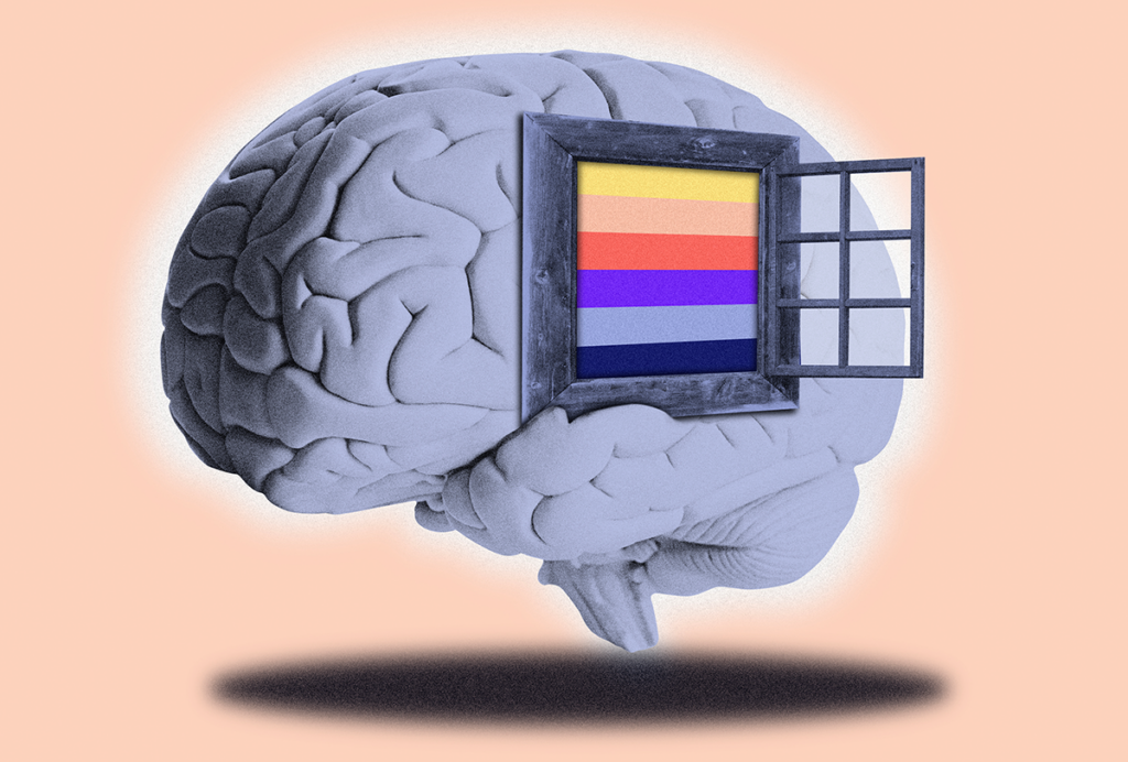
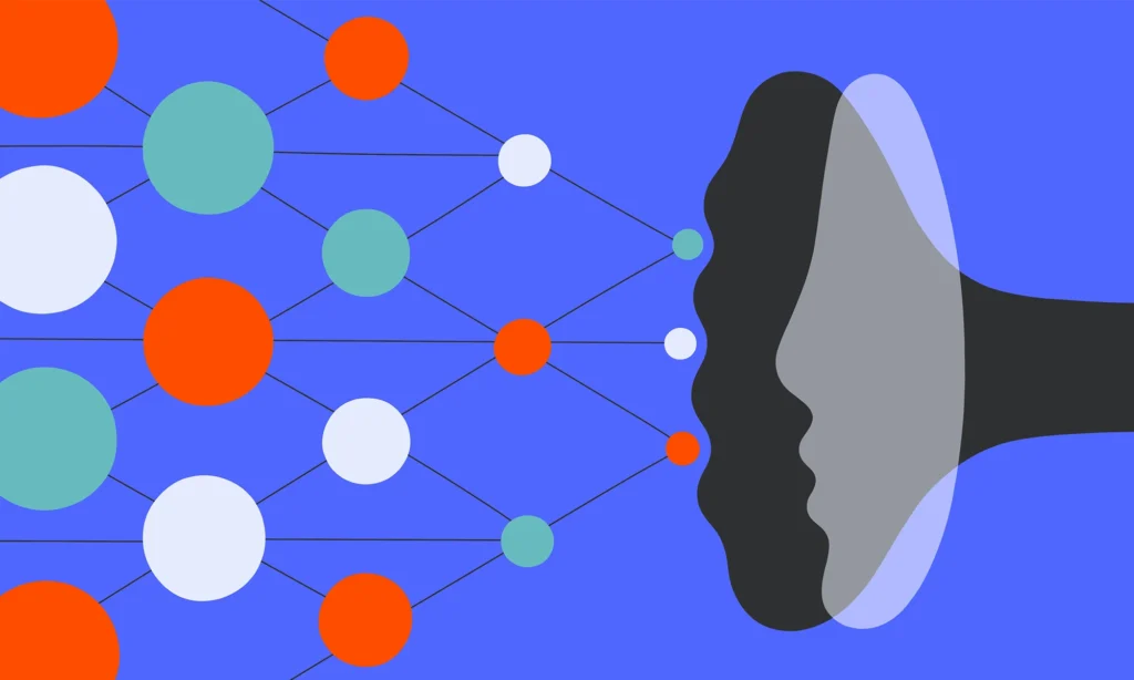
Human thought and behavior emerge through complex and reciprocal interactions that link microscale molecular and cellular processes with macroscale functional patterns. Functional MRI (fMRI), one of the most common methods for studying the human brain, detects these latter patterns through the “blood oxygen level dependent,” or BOLD, signal, a composite measure of both neural and vascular signals that reflects an indirect measure of brain activity. Despite an enormous investment by scientific funders and the research community in the use of fMRI, though, researchers still don’t fully understand the underlying mechanisms that drive individual or population-level differences measured via in-vivo brain imaging, which limits our ability to interpret those data.
For fMRI to meaningfully contribute to progress in neuroscience, we need to develop research programs that link phenomena across levels, from genes and molecules to cells, circuits, networks and behavior. Without a concerted effort in this direction, fMRI will remain a methodological spandrel, a byproduct of technological development rather than a tool explicitly designed to reveal neural mechanisms, generating isolated datapoints that are left unintegrated with broader scientific theory or progress.
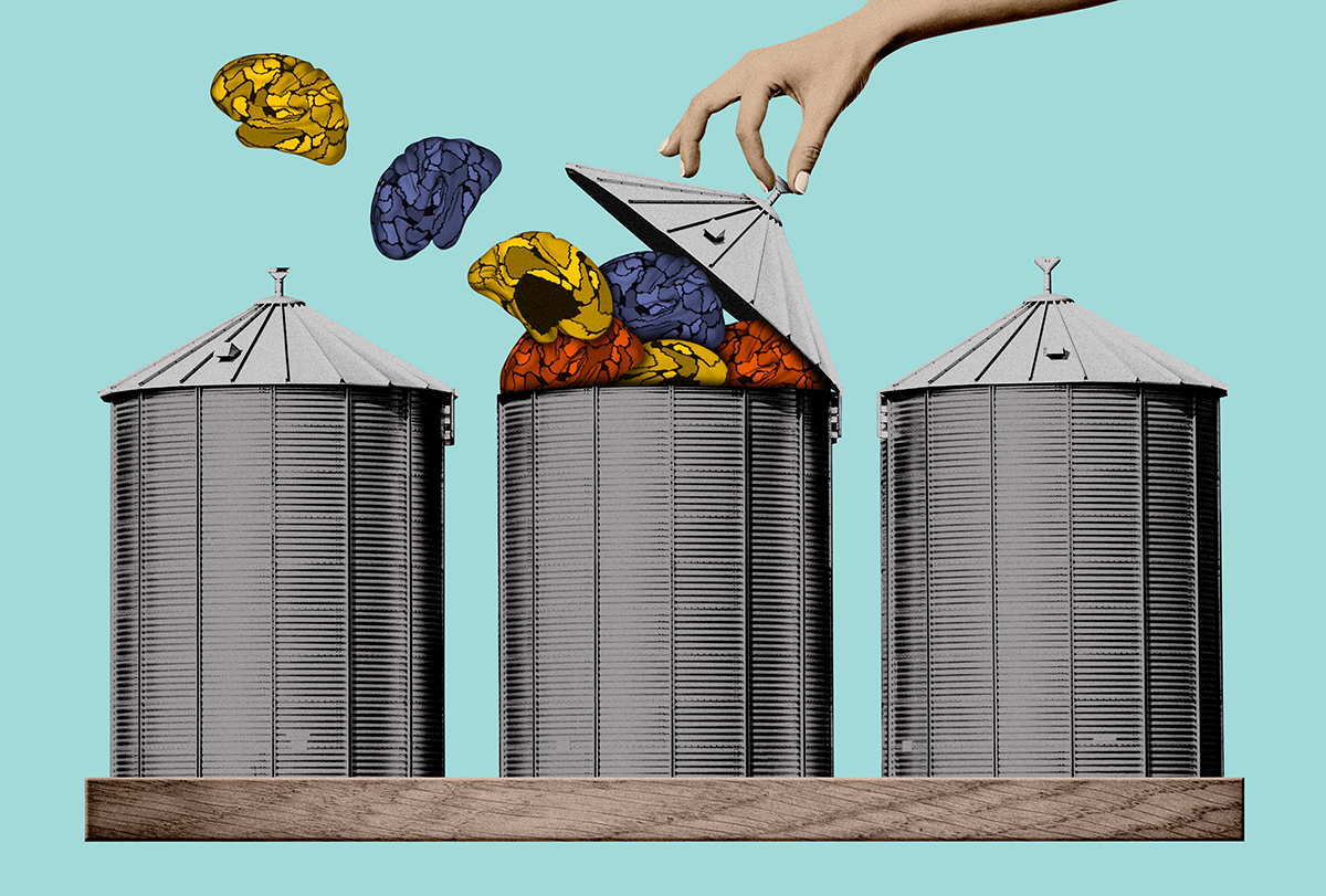
Recently, the human functional neuroimaging community has turned a critical eye toward its own methods and findings. These debates have led to field-wide initiatives calling for larger and more diverse study samples, better phenotypic reliability and findings that generalize across populations. But researchers have put relatively little emphasis on contextualizing the resulting work across levels of analysis or on deciphering the biological mechanism that may underpin changes to the BOLD signal across groups and individual people or over the lifespan.
Appeals to better integrate the different levels of neuroscience are not new. But despite persuasive arguments, fMRI researchers have largely remained scientifically siloed, isolated by a nearly ubiquitous focus on a single level of analysis and a rigid adherence to a select set of imaging methods. Our work is typically presented inside of field-specific echo chambers—departmental or group seminars, topic-specific journals and society meetings—where our methodological and analytic choices go unchallenged. What progress can we expect to make if we remain isolated from other fields of study?
True scientific advancement will require adopting novel methods and integrating findings from allied disciplines. The PsychENCODE initiative in genetics offers a powerful example of the progress that is possible through multidisciplinary collaborative team science. Until recently, in-vivo brain imaging has largely been absent from these kinds of multidisciplinary and integrative efforts, limiting the opportunities to contribute our discoveries, theories or vision toward a comprehensive view of brain function.
Fortunately, a cultural revolution is on the horizon. A new generation of researchers is driving a sea change in the way data are collected and shared across the sciences. Historically, linking levels of analysis has been difficult, if not impossible, to accomplish, because individual research faculty were incentivized to retain tight control over their data. This has changed dramatically in the past 10 to 15 years with the explosion of open science initiatives, which now make it possible to combine data from previously distinct areas of neuroscientific inquiry.
F
or example, there has been an exponential increase in the availability of imaging genetic and population neuroscience datasets, in which we have both structural genetic data and MRI-based measures of brain structure and function from tens of thousands of people. In parallel, open-access postmortem tissue repositories have increased in both number and kind, with datasets including the Allen Human Brain Atlas, BrainSpan and NIH Genotype-Tissue Expression now providing data from hundreds of tissue samples across the lifespan, often with regional labels or reference coordinates common to fMRI datasets (e.g., MNI space).As a result, a diverse set of research groups have begun to mine these data, integrating them with population-level cohorts that encompass in-vivo measures of brain structure and function, as well as multiple domains of cognition, behavior and genetics. The resulting bulk consolidation and open dissemination of these diverse initiatives provides researchers seeking to bridge disciplinary boundaries an extraordinary opportunity to tackle questions at the intersections of traditionally distinct fields of study.
Researchers in our laboratory, as one example, have worked to integrate spatial variations in postmortem gene-expression and cell-type distributions with brain networks identified via fMRI. These data have revealed that the in-vivo functional organizational properties of the brain are reflected in the topography of gene transcription. Researchers can now nominate cell types involved in specific diseases based on the circuits implicated in brain imaging studies. Recently, we have expanded this work, demonstrating that cell-type distributions are spatially coupled to the functional organization of cortex, as estimated through intrinsic (resting-state) fMRI. These relationships are so robust that researchers can now look at the cell-type distributions in a postmortem brain tissue sample and predict what functional network the sample came from.
Our work in this space reflects a small piece of the converging evidence establishing how microscale features of neurobiology, including receptor densities, gene expression and patterns of cellular distribution, may underpin the in-vivo functional organization of the human brain as assessed through fMRI. It is hard to overstate the potential utility of these multiscale approaches; they provide an empirical pathway to establish the molecular and cellular mechanisms underlying the patterns we measure with fMRI and may lead to new clinical applications.
How as a field can we incentivize cross-field multiscale research programs? Here I outline a few recommendations, drawn largely from my own experiences and conversations with collaborators and colleagues over the years.
First, we need to actively stamp out “paradigm wars” that can often arise as a byproduct of the methodological dogmatism rampant amongst scientists. This form of gatekeeping is akin to the famous “no-true Scotsman” logical fallacy—in this case, “no true neuroscientist studies X at level Y.” When our fields of study are reified—viewed as unitary, enduring and fully isolable from other areas of science—research that bridges departments is minimized or lost to misguided appeals for scientific or methodological purity. Second, our coursework and training programs must foster appreciation of diverse methods and theoretical approaches. This can be addressed, at least in part, through the inclusion of cross-departmental course offerings and seminars, interdigitated lab spaces, and institutes that house multiple academic departments. Third, scientific funders need to prioritize support for cross-field programs of research. To do so, they must construct study sections whose members appreciate the importance of methods and approaches outside of their own field of study.
Finally, the scientific community needs to develop processes to support, hire and retain researchers whose work sits between traditional fields of study. Here, academic performance metrics are often tailored to traditional area boundaries—that will need to change. It will be critical for us as individual scientists to push back against siloed beliefs, take an honest look at our field’s methods, their potential for true scientific discovery and progress, and the likely benefits for integrating approaches across disciplines.