Timing tweak turns trashed fMRI scans into treasure
Leveraging start-up “dummy scans,” which are typically discarded in imaging analyses, can shorten an experiment’s length and make data collection more efficient, a new study reveals.
For functional MRI (fMRI) studies, time is money.
“You want to get the most bang for your buck,” because using a scanner is expensive, says Lucina Uddin, professor of psychiatry and biobehavioral sciences at the University of California, Los Angeles.
Yet, for every experiment, most scientists throw out the first 10 to 20 scans they capture. These “dummy scans” are typically not analyzed because they are collected before the machine’s applied magnetic field—which is generated by a radio frequency (RF) pulse—has reached a steady state, Uddin says.
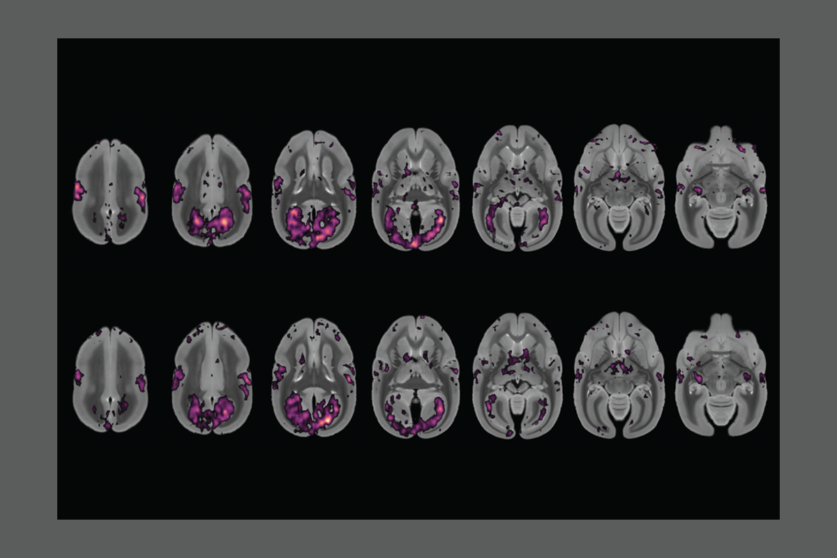
But that start-up period can actually offer some of the best data a scanner produces, a new preprint suggests. By acquiring event-related scans as the field ramps up and correcting for the changing baseline signal, a team can run half as many trials and still reach the same statistical threshold, the study reports.
The new approach provides a simple and accessible way for imaging researchers to improve the signal-to-noise ratio of their data in certain circumstances, says Peter Bandettini, chief of the Section on Functional Imaging Methods and director of the Functional MRI Core Facility at the National Institute of Mental Health, who was not involved in the work.
“That should have been tried years ago,” he says. “It shows that there’s still tricks that you can play with the scanner and acquisition to get something out of it.”
T
he findings came about almost by accident, says Ravi Menon, professor of medical biophysics and medical imaging at the University of Western Ontario, who led the new work.His team wanted to collect fMRI and electrophysiological data simultaneously from marmosets, but the magnetic field changes generated by the scanner’s RF pulses interfere with the electrical signals, he says. The pulses shift the magnetization of brain tissue, making it detectable by RF coils that are used for imaging.
Menon and his team decided they could use quiet, pulse-free periods to collect their electrophysiological data, as some past studies have done to help participants in the scanner hear audio stimuli. But those paradigms were set up to collect data at regular, short intervals, and the team wanted to be able to trigger data collection at any moment in response to a stimulus, says study investigator Renil Mathew, a graduate student in Menon’s lab.
So the team presented an auditory tone or a flickering light to a head-fixed marmoset in a 9.4 tesla scanner and then generated RF pulses and collected data for 12 or 16 seconds, respectively. They alternated this block of scans with an equal-length block of task-free scans, which enabled them to obtain a signal baseline, and interleaved those blocks with an 8-second “acquisition-free period.” They repeated this entire process 14 more times.
Extending the time between pulses lengthens the overall scan and can introduce noise from background tissue, Menon says. But protons in brain tissue most closely align their spin to the RF-generated magnetic field—and thus generate their strongest signal—with the first pulses; if the pulses come in quick succession, the particles begin to respond less vigorously.
Typical fMRI experiments acquire their scans during that low-response steady-state period, when changes in oxygenation, which give rise to the BOLD signal, are thought to be most easily detected. But because the team presented their stimuli before starting the RF pulses, the stimulus-driven BOLD signal—which is delayed relative to electrophysiological measures—arrived during the high-signal period, leading to better BOLD detection. By waiting long enough in between scan acquisitions, they could harness that higher sensitivity repeatedly.
“Rather than using heavily computational models to clean the data, we just use basic physics to get around that problem,” Mathew says. The approach roughly doubled the team’s ability to detect a signal over time, he and his colleagues found.
Although the paradigm came about to help collect simultaneous electrophysiological data, “it started to become evident that we actually have something with higher sensitivities” that could also be useful for other experiments, says study investigator Amr Eed, a postdoctoral researcher in Menon’s lab. They posted the findings on bioRxiv last month.
T
he approach is not going to work with every experiment, Uddin cautions. “It’s a great proof of concept. The question is going to be, how translatable will this be to human neuroimaging, human fMRI and cognitive neuroscience?”The approach is well suited for timing data collection to a particular event, including collecting scans immediately after EEG detects a seizure, which is something the team plans to do in the future, Menon says. And although the experiments were run using event-related fMRI, they could also be adapted for collecting resting-state fMRI, Mathew says.
For situations in which the physiological noise is greater than that induced by the scanner, the paradigm is unlikely to offer any advantage, Bandettini says. But “in those cases where you’re really pushing the resolution, like with layer fMRI in humans, then this technique might be worth trying.” Establishing a parameter space in which this approach is useful would be helpful for the field, he adds.
The biggest advantage may be for animal behavioral experiments, which are mostly done on scanners with high field strengths, Eed says. He adds that he and his colleagues next plan to move forward with their experiments collecting simultaneous fMRI and electrophysiological data to help localize neural activity.
“You’re getting two different modalities in a clean way, which we never used to get before,” Mathew says. And as a bonus, it does not interfere with the fMRI data, he adds. “It enhances it.”
Recommended reading
Breaking down the winner’s curse: Lessons from brain-wide association studies
Split gene therapy delivers promise in mice modeling Dravet syndrome
U.S. human data repositories ‘under review’ for gender identity descriptors
Explore more from The Transmitter
