Digitization of ‘breathtaking’ neuroanatomy slide collection offers untapped research gold mine

Thousands of histological sections of vertebrate brains—including from spiny dogfish, turtles and more—are newly available online.

By Shaena Montanari
22 January 2025 | 3 min read
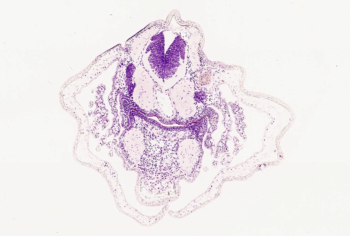
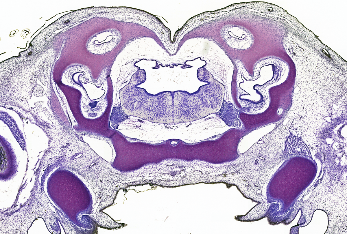
Violet-stained neurons in a serial transverse section from an embryonic spiny dogfish, a bottom-dwelling shark.
Baby shark: Violet-stained neurons pepper serial transverse sections from an embryonic spiny dogfish (Squalus acanthias), a bottom-dwelling shark (MCZ:SC:3780, slide 12). This slide is one of 48 from the embryo.
R. Glenn Northcutt Collection of Comparative Vertebrate Neuroanatomy and Embryology

For evolutionary neuroanatomists who compare diverse animal brains, access to a gold mine of 500,000 histological sections and whole mounts is now only a mouse-click away.

The R. Glenn Northcutt Collection of Comparative Vertebrate Neuroanatomy and Embryology at Harvard University—which comprises 33,000 slides of tissue samples from more than 240 vertebrate genera—is one of the world’s largest and most diverse collections of its kind. Northcutt, a prolific comparative vertebrate neuroanatomist and emeritus professor of neurosciences at the University of California, San Diego, amassed the collection over the course of five decades. Since 2021, James Hanken, research professor of biology at Harvard University and curator at the Museum of Comparative Zoology, has led an effort to digitize it.

The scanning process is still ongoing and may take another two years to complete, Hanken says, but more than 8,000 slides are already publicly available in two online data repositories: MCZBase and MorphoSource. 

A comprehensive inventory of the entire collection appears in a paper Hanken and his colleagues published last week in the Bulletin of the Museum of Comparative Zoology. It provides researchers with an in-depth guide for using the collection, Hanken says.

Few other resources of this type are available online to researchers interested in evolutionary biology and brain anatomy, says Andrew Iwaniuk, professor of neuroscience at the University of Lethbridge. For example, neither the Welker Comparative Anatomy Collection nor the Starr Collection, both housed at the U.S. National Museum of Health and Medicine in Silver Spring, Maryland, are available online. To access slide collections such as these, scientists have had to travel to see them in person, which can be difficult for those outside the United States, Iwaniuk adds.

“These days, there’s more and more people [who] are interested in these big evolutionary questions when it comes to the nervous system,” he says. “Those sorts of questions can really only be best answered by having these broad comparative anatomical collections that take a lifetime to assemble.”
research image of a serial transverse section from a tuatara Sphenodon punctatus
research image of peripheral nerves stained violet in a transverse section of a whole embryo of the endangered Australian lungfish
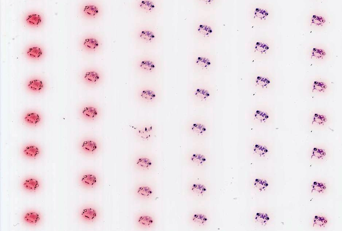
Research image of histological sections of channel catfish embryos
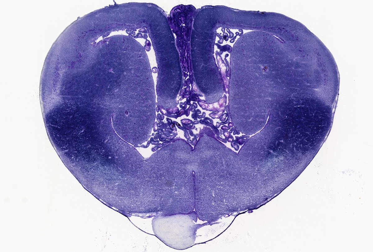
Research image of a histological sections of a painted turtle
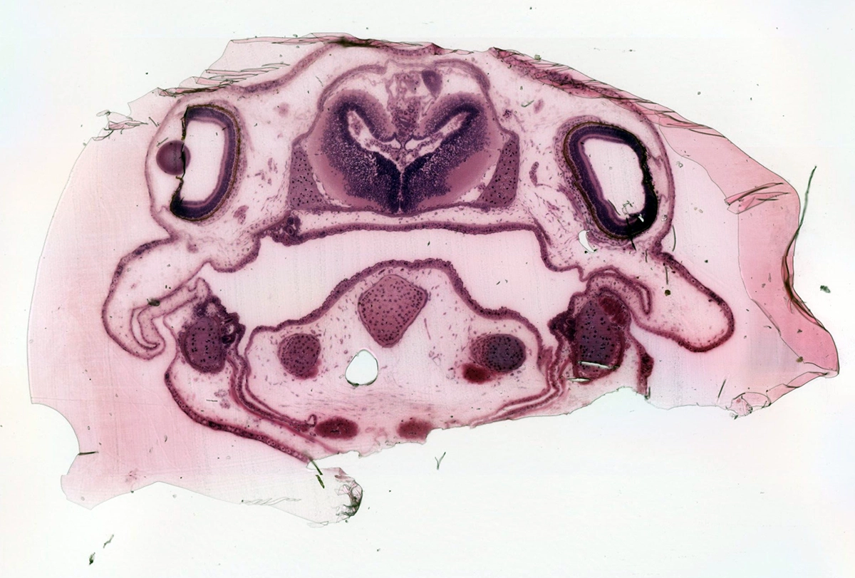
Research image of a transverse section of the head of a juvenile axolotls Ambystoma mexicanum
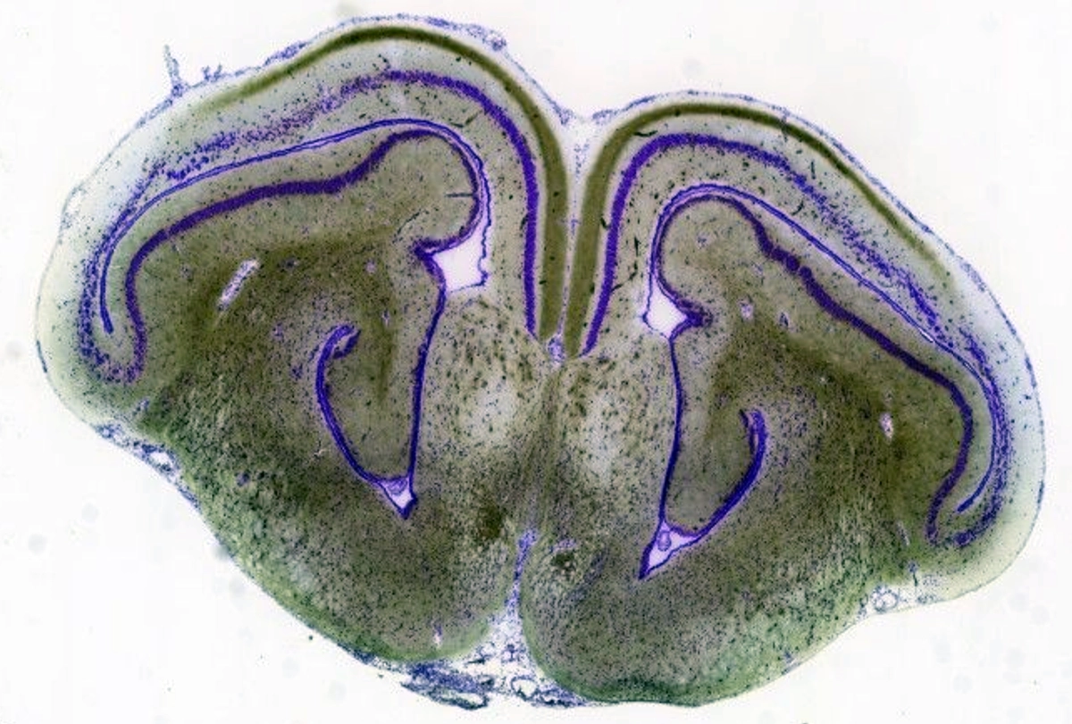
Sole survivor: Brown staining tracks acetylcholinesterase, a marker of cholinergic neurons, through a serial transverse section from a tuatara (Sphenodon punctatus), a rare and endangered reptile from New Zealand that is the last living species of its order. (MCZ:SC:4050, slide 15)
R. Glenn Northcutt Collection of Comparative Vertebrate Neuroanatomy and Embryology

N

orthcutt published hundreds of comparative neuroanatomy papers after viewing the slide collection under a microscope. He described the emergence of the mammalian neocortex and how the neural crest led to the origins of vertebrates, among many other discoveries.

Many more research papers stand to be unearthed from this resource now that it has gone digital, Hanken says. “The number of non-model species available for doing comparative studies is breathtaking.”

Once digitization is complete, the online resources promise to make 270 species available for viewing, including unusual entries such as numerous lungfish species, coelacanths and tuataras. There are also representative samples from amphibian, marsupial and primate species.

Even though slides may seem like a museum relic compared with MRI and CT scanning, new technologies still cannot quantify neuron numbers in most vertebrate brains, Iwaniuk says. “So the fallback will be histology for the foreseeable future of comparative and evolutionary neuroscience.”

Sign up for our weekly newsletter.

Catch up on what you may have missed from our recent coverage.