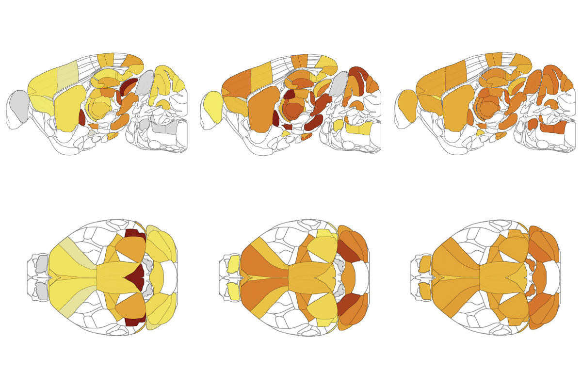
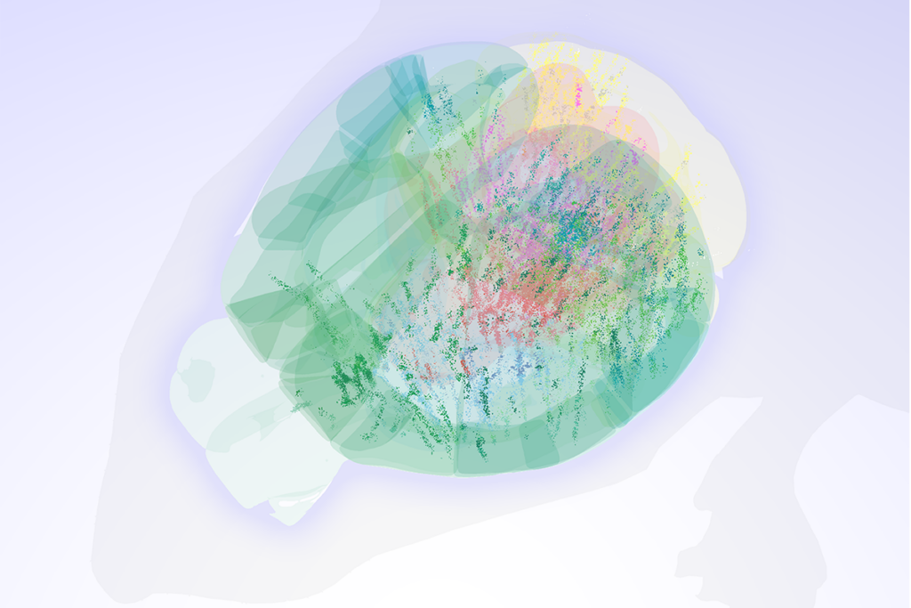
Choosing what ice cream to buy may take only a couple of seconds. But in the moments between seeing dozens of flavors and selecting chocolate, a flurry of activity courses through a person’s brain. This decision-making surge flows across multiple brain areas in parallel as information guiding the choice—the taste or color of each flavor—accumulates, two new independent studies in rodents suggest.
Both teams recorded from widespread areas across the brains of either head-fixed or freely moving rodents as the animals performed decision-making tasks. As a result, the findings give a detailed view of how widely distributed the activity underlying decision-making is, says Daeyeol Lee, Bloomberg distinguished professor of neuroscience at Johns Hopkins University, who was not involved in the new studies. This insight expands the field’s understanding of this process beyond the frontal cortex, striatum and superior colliculus, which were implicated in past studies that looked only “at snapshots of a very complex system.”
The new work also helps scientists disentangle whether the signals they previously picked up in freely moving animals were due to the abstract nature of the decision-making, the animals’ movement, or both, says Matteo Carandini, professor of visual neuroscience at University College London, who was not involved in the new work. “When you’re trying to understand an abstract decision, because there’s so much embodiment of decisions, that really poses a problem.”
The cells that respond to sensory information overlap with those that help plan a movement, one of the studies revealed. And cells across the brain coordinate their activity right up until an animal settles on its pick, according to the other study.
That synchronization ensures that the same information is transmitted across different parts of the brain, and it also helps the animal effectively act on the selection, says Thomas Mrsic-Flogel, professor of neuroscience at University College London, who led one of the studies. “These dimensions of activity that integrate the sensory input, or the ones that prepare the movement—they’re all coordinated between those regions.”
M
ice viewed black and white parallel lines drifting across a monitor and learned to remain stationary until the lines changed speed. This stimulus initially activated neurons in a restricted set of brain areas—including the visual cortex and the superior colliculus, Mrsic-Flogel and his colleagues found when they recorded from around about 15,000 single cells across 51 brain regions.But then the neuronal activity expanded beyond those areas. Neurons in multiple brain regions downstream of the visual system, including those in the frontal motor cortex, thalamus and basal ganglia, encoded increasing levels of information about the video, the team found. The building evidence about the lines’ velocity told the mice when they had seen enough to be confident that the speed had changed and to therefore move their body to report their decision, Mrsic-Flogel says.
The preparatory buildup of neuronal activity before a decision, also seen in previous studies, correlates and overlaps with the activity of the neurons that integrate sensory evidence—but not with activity of cells that control the movement itself, Mrsic-Flogel and his team found. The preparatory and evidence activity “appear to be the same kind of signals in the brain,” Lee says.
That overlap makes sense as a way for the mouse’s brain to convey accumulated sensory information to the parts of the brain responsible for acting on its choice, Mrsic-Flogel says.
In untrained mice, on the other hand, the sensory evidence-associated activity was present in the visual system but did not spread to other regions, such as the striatum. That suggests that the widespread distribution of the activity is enabled by learning the task, Mrsic-Flogel says. If the mouse has not learned, he adds, “it doesn’t care; it doesn’t accumulate the evidence.” He and his colleagues reported their findings in Nature last month.
T
he widespread neuronal activity involved in decision-making is also tightly coordinated across some of the same dispersed brain areas, according to the other study, which was posted on bioRxiv in August.In this work, rats learned to tally clicks played from speakers on their left and right sides and then nose-poke a hole on the side that put out more clicks. A frontostriatal subnetwork—formed by the primary motor cortex, the dorsomedial prefrontal cortex and the anterior dorsal striatum—drove the evidence accumulation and decision-making in the animals, the team found by simultaneously recording from thousands of neurons via eight Neuropixels probes in each animal.
The activity representing sensory evidence in each of these areas also increasingly predicted the animal’s ultimate choice—but only up until a certain point, the team found. That point, which the team previously dubbed the “neurally inferred time of commitment,” indicates when an animal has formed a decision. It happens at different times during each trial and in each animal, and it is not time-locked to either the external stimulus or the movement, the team discovered.
Once the animal commits to a choice, a brain-wide shift occurs, the team found. Neuronal firing rate patterns change, the strong correlations in brain activity across regions disappear, and the models’ ability to predict an animal’s choice from its neural activity stops improving. It seems that, after this time point, the brain stops integrating evidence for the decision, says Carlos Brody, professor of neuroscience at the Princeton Neuroscience Institute, who led the work. “We were just gobsmacked to see this.”
Although the biomarker for the time of commitment shows up throughout the brain, it appears earliest in the primary motor cortex, the team also found. This area may be where the process of forming a decision ends and executing it begins, Brody says.
Similarly, Mrsic-Flogel and his colleagues identified that the neurons that integrate evidence in specific brain regions—such as the frontal cortex, basal ganglia, cerebellum and midbrain—are also the ones that come online first, before an animal makes its decision. For committing to a decision, “those might be the most important ones,” he says.
B
oth new studies made use of a massive number of recordings, Carandini says, noting that the work from Brody and his colleagues “pushes the frontier by recording all of those neurons at the same time.” But that team recorded from only three rat brains—and the rodents may have made slight movements unseen by the scientists during the trial, which could have masked the purely decision-making signals, he says. “You don’t know if [the rodent] is moving the tail a little bit.”And the studies did not answer which specific sites within brain regions drive evidence integration, Mrsic-Flogel acknowledges. “Let’s say we find that neurons have integrated evidence in the striatum, which people have also found before. Which division of the striatum is it?”
These findings in rodents cannot tell us everything about the neural mechanisms of human decision-making during complex cognitive tasks, Lee says. But for simple decisions, such as whether a stimulus falls on one side or another, the mechanisms might hold up, Carandini says. “I would be shocked if the brain of a human solved it in a different way.”


