Timothy Roberts is Oberkircher Family Chair in Pediatric Radiology and vice-chair of radiology research at the Children’s Hospital of Philadelphia. He is also professor of radiology at the University of Pennsylvania.
Timothy Roberts
Professor of radiology
Children's Hospital of Philadelphia
From this contributor
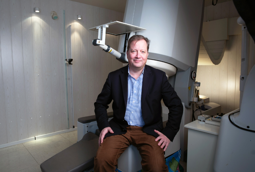
In quest for autism biomarkers, this technique has magnetic appeal
To find biological markers of autism, scientists would be wise to measure the brain's electrical activity along with the resulting magnetic fields.
In quest for autism biomarkers, this technique has magnetic appeal
By
Timothy Roberts
26 March 2019 | 6 min read

When tracking brain activity, timing can be key
A brain imaging technique called magnetoencephalography characterizes not just what is happening in the brain, but also where and when, making it ideally suited for studying autism.
When tracking brain activity, timing can be key
By
Timothy Roberts
15 March 2016 | 5 min read
Imaging biomarkers could signal autism spectrum disorder
There does not appear to be a single genetic or environmental cause of autism, and given the heterogeneity of symptoms, coming up with a clear yes or no test for autism is challenging. Timothy Roberts argues that imaging and electrophysiology are key in the search for autism biomarkers.
Imaging biomarkers could signal autism spectrum disorder
By
Timothy Roberts
12 April 2011 | 4 min read
Explore more from The Transmitter
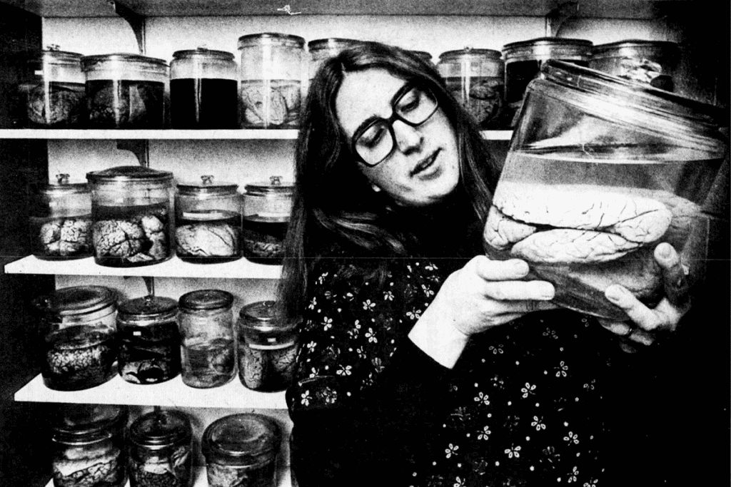
Remembering Barbara Finlay, ‘maverick’ of evolutionary neuroscience
Finlay, who died on 12 September, championed the idea that developmental constraints dictate how evolution sculpts brains, a concept she used to model the timing of hundreds of events and align them across mammalian species.
Remembering Barbara Finlay, ‘maverick’ of evolutionary neuroscience
Finlay, who died on 12 September, championed the idea that developmental constraints dictate how evolution sculpts brains, a concept she used to model the timing of hundreds of events and align them across mammalian species.
By
Calli McMurray
23 October 2026 | 8 min read
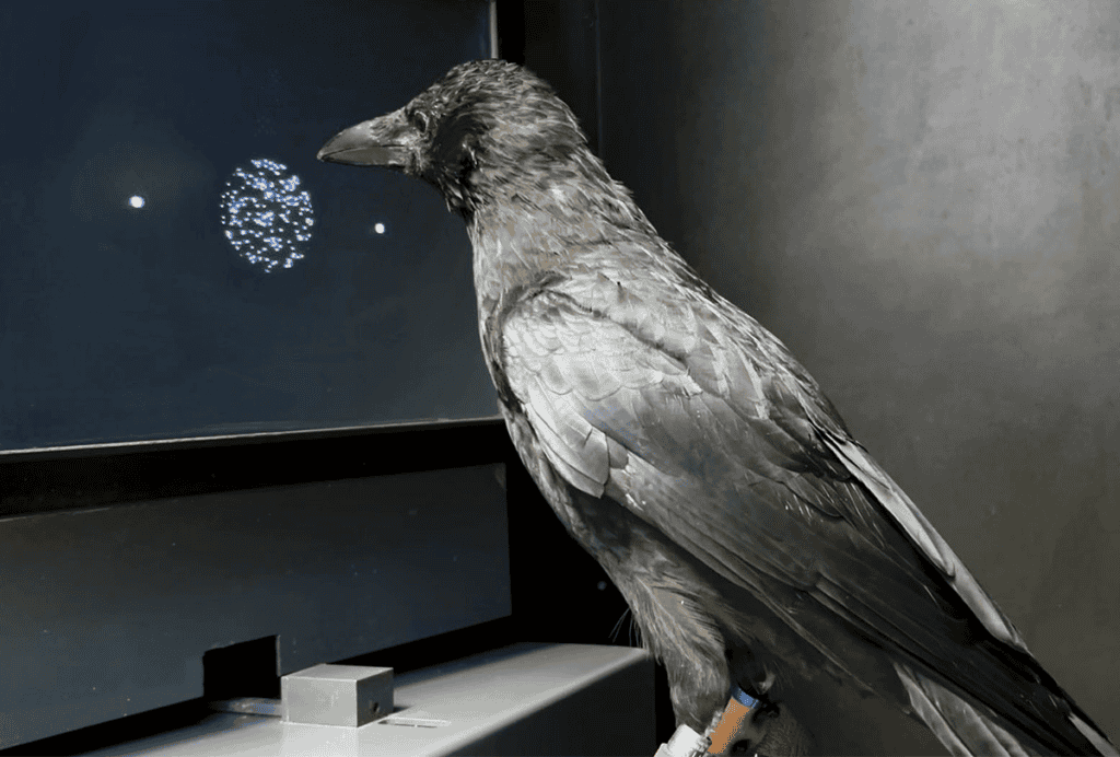
Crows have brains ‘in the fast lane’
The birds accurately complete decision-making tasks twice as fast as primates, although their endbrains look considerably different.
Crows have brains ‘in the fast lane’
The birds accurately complete decision-making tasks twice as fast as primates, although their endbrains look considerably different.
By
Sarah Thau
22 October 2026 | 4 min read

Prevalence of profound autism declines with new definition
The updated, more precise definition used in a CDC study includes adaptive scores.
Prevalence of profound autism declines with new definition
The updated, more precise definition used in a CDC study includes adaptive scores.
By
Brianna Abbott
22 October 2026 | 5 min read