When John Tuthill was a postdoctoral researcher at Harvard Medical School, he worked just down the hall from Wei-Chung Allen Lee, who was developing new technology to image and map cell connections in the central nervous system. Lee wanted to use his technique in the fruit fly Drosophila, but he knew that other groups were already making such images of the fly brain. So Tuthill, who was studying touch stimuli in Drosophila, suggested Lee pivot to map the fly’s ventral nerve cord (VNC) instead.
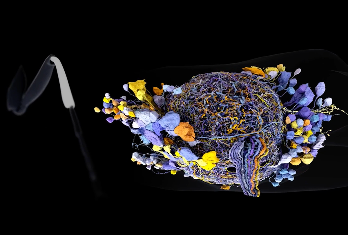
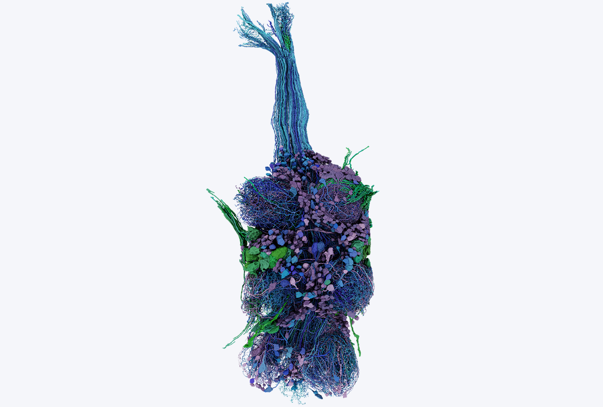
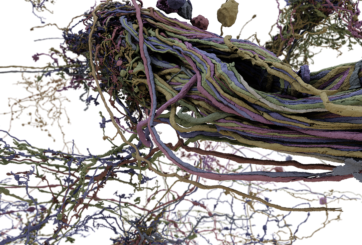
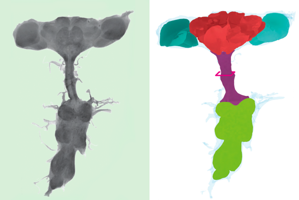
A decade later, Tuthill, Lee and colleagues have published a map of the connections among motor neurons in a female fly’s VNC, which is analogous to the spinal cord in mammals. The diagram, published on 26 June in Nature, details roughly 45 million synapses that connect nearly 15,000 neurons, and is the second such connectome to be released. A different team, at Howard Hughes Medical Institute’s Janelia Research Campus, published a male fly’s VNC connectome to eLife’s preprint server in June 2023. (The team posted an updated, reviewed preprint on 23 May 2024.)
“The connectome is only useful if you can connect it to the muscles,” says Tuthill, associate professor of neuroscience at the University of Washington. “The output of the connectome is the activity of motor neurons.”
With connectomes from both a male and a female fly, researchers are starting to look for differences not only between individuals, but between the sexes. An initial comparison of the two connectomes, posted to bioRxiv on 28 June by members of both teams, including Tuthill and Lee, identified circuits that appear to control sex-specific behaviors, including male courtship songs and the female extension of an organ used to deposit eggs.
For decades, neuroscientists have dreamt of complete wiring diagrams of the brain, home of such complex and alluring processes as decision-making, abstract thinking and consciousness. Last August, the FlyWire Consortium based at Princeton University revealed the first full fruit fly connectome, containing nearly 130,000 neurons.
The VNC has drawn less attention, Tuthill says, in part because “it seems a little less magical.” But the nerve cord is the direct connection to what an animal actually does: move.
“It’s where the rubber meets the road,” Tuthill says.
T
he teams behind both connectomes, often referred to as the female adult nerve cord (FANC) and male adult nerve cord (MANC), followed broadly the same process: Algorithms made an initial map and people proofread the computer’s work.For the MANC, however, the Janelia team hired and trained proofreaders, keeping the data private until the map was complete. By contrast, the FANC team adopted FlyWire’s open-source, community-driven approach. Researchers in the fly-brain community voluntarily proofread the map. The result is a constantly evolving dataset, with varying stages of completeness and quality for different parts of the nerve cord.
Tuthill and his colleagues combined imaging data made using Lee’s technology, known as GridTape, with neural images made using an electromagnetic-radiation technique called X-ray holographic nanotomography.
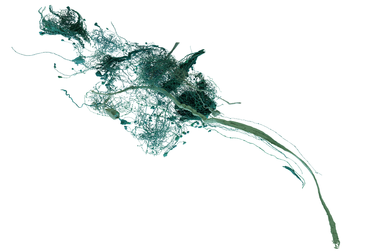
After identifying 17,076 cells in the VNC, of which 14,621 are neurons, the researchers mapped connections between the motor neurons, among the largest cells in the fly’s nervous system. (The largest motor neuron, the fast tibia flexor, has dendrites that total nearly 1 centimeter in length, despite the average fly being only 3 millimeters long.)
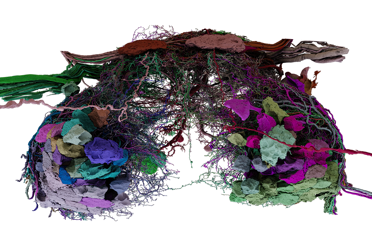
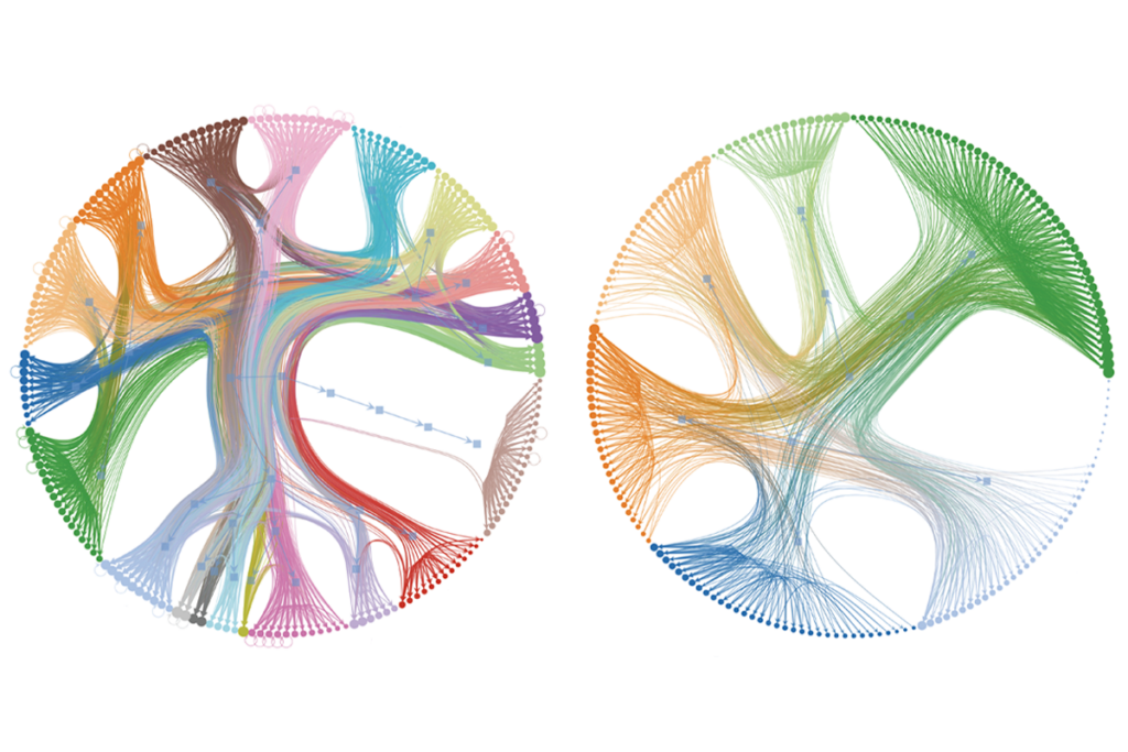
The resulting map details 69 leg motor neurons that are connected to 1,546 pre-motor neurons via 212,190 synapses, as well as 29 wing and thorax motor neurons with 144,668 synapses connecting them to 1,784 pre-motor neurons.
The motor neurons that control the fly’s legs are grouped into “modules” that are all controlled together, according to a companion paper the team also published in Nature. This module design, which also applies to the motor neurons that control the wings, frees the brain from needing to send signals to each neuron individually, Tuthill says.
