Listen to this story:
0:00
/
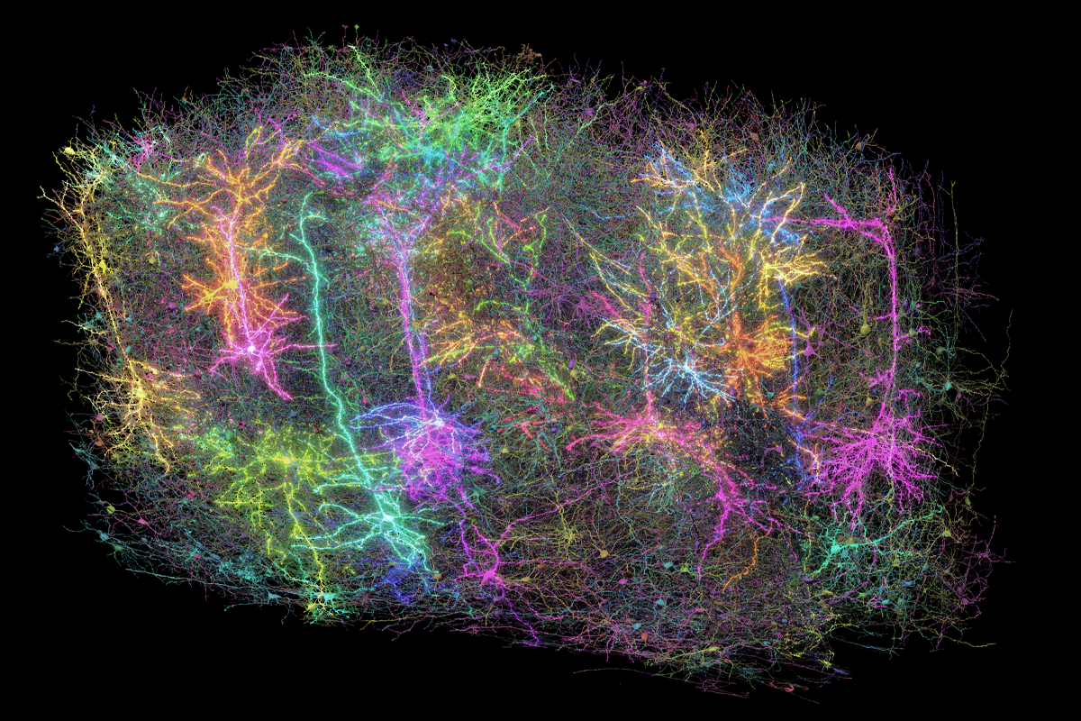
A cubic millimeter of brain tissue, meticulously sectioned, stained and scrutinized over the past seven years, reveals in stunning detail the role of inhibitory interneurons in brain structure and function.
The most comprehensive map to date of cell structure and function in the mouse cortex reveals a previously unappreciated level of coordination among inhibitory interneurons.
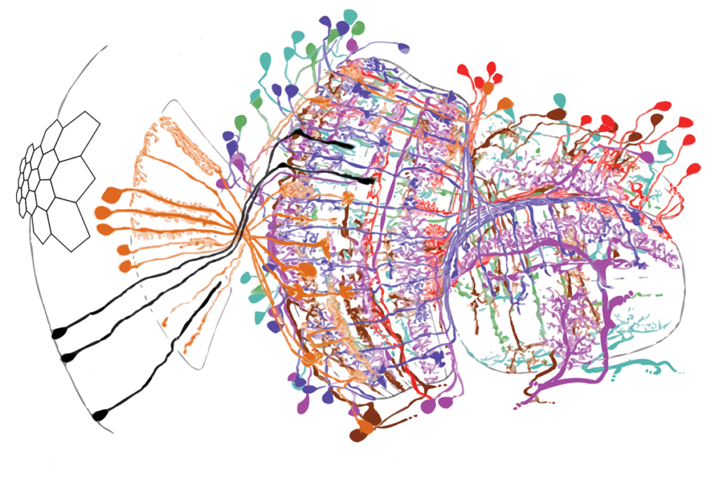
The study—one of 10 on the mouse connectome released today in the Nature family of journals—shows that interneurons carefully select the types of excitatory neurons they connect with. They also appear to work in teams, targeting the same type or types of excitatory neurons from different angles.
“Not only is there this remarkable specificity of inhibitory cells to a particular set of excitatory types, but even inhibitory cells that come from very different groups can share that specificity,” says Nuno Maçarico da Costa, associate investigator at the Allen Institute, who led the interneuron study.
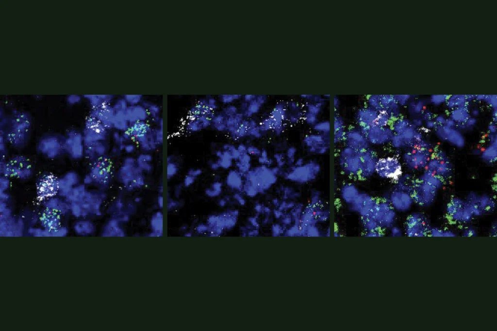
To build the map, researchers recorded neuronal firing in a cubic micrometer of visual cortex as a mouse watched a video and ran on a treadmill. They then used electron microscopy to trace the 1,183 excitatory neurons, 164 inhibitory interneurons and more than 70,000 synapses that orchestrated the bursts of brain activity.
The interneuron analysis adds to mounting evidence that inhibitory cells working in concert are the true maestros of brain activity.
“I think the hints of a lot of what they’re saying have been coming in multiple ways, but the ability to get down to the ultrastructural level and look at it has been fantastic,” says Gord Fishell, professor of neurobiology at Harvard Medical School, who was not involved in the work.
Drugs that target different teams of interneurons could potentially turn up or down the volume of specific neural circuits, says study investigator Clay Reid, senior investigator at the Allen Institute and a lead on the Machine Intelligence From Cortical Networks (MICrONS) project.
“Without understanding the structure of the cortical circuit, it’s very difficult to understand the influence of current and certainly future medications on that circuit,” Reid says. Now, “one of the knobs that you can turn medically is far better understood.”
Fishell agrees, and he likens efforts to “turn on” certain neurons to “playing a piccolo in the middle of a concert and thinking you’re going to make it sound better.” A better goal is to “gain control” of the symphony, he says, “because the biggest problem with brain function is the lack of synchronization and lack of coordination.”
T
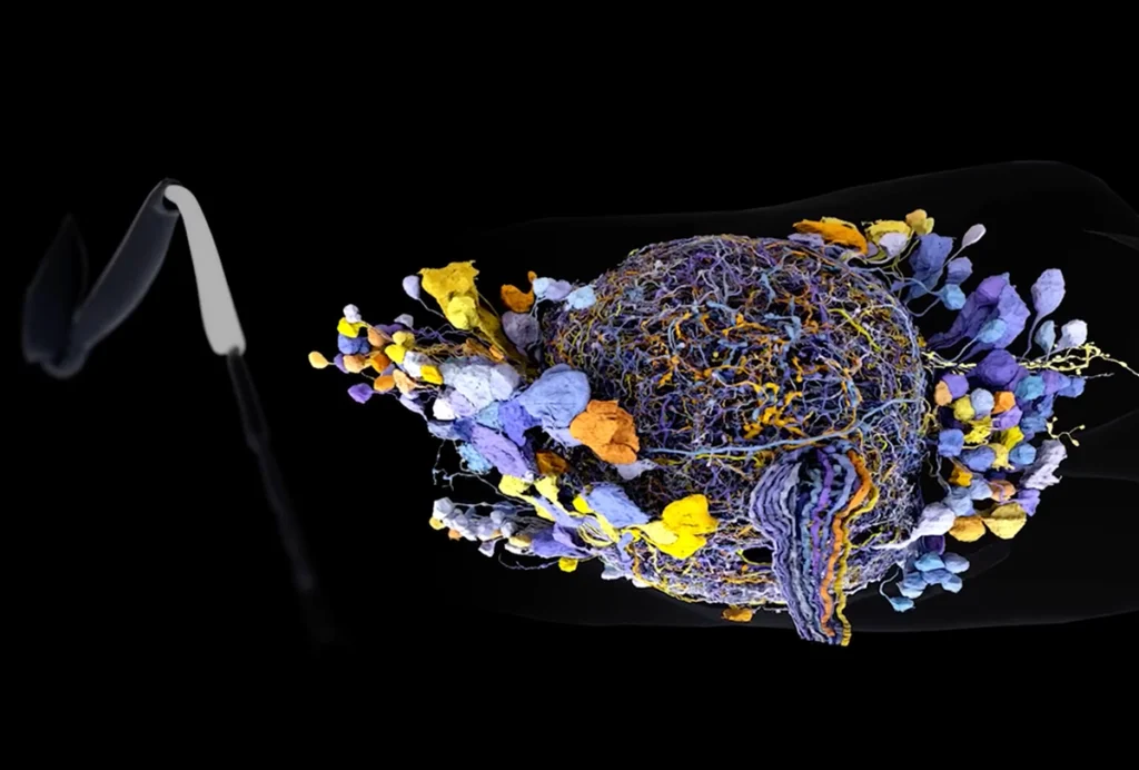
he collection of 10 papers is the culmination of seven years of work by more than 150 scientists. One of the papers describes the 1.6 petabytes of detailed structural and functional data for a cubic millimeter of mouse cortex. The researchers leveraged a mix of human skill, machine learning and artificial intelligence to analyze more than 95 million electron micrographs. The data are public in the form of an interactive online resource called the MICrONS Explorer.“You start with humans manually painting, and then you need to bootstrap using machine learning to be able to run across the whole dataset,” says study investigator Forrest Collman, associate director of data and technology at the Allen Institute.
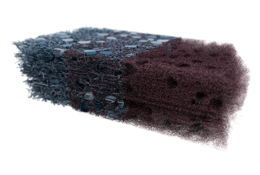
The result is a map with an “exquisite” level of detail that researchers can use to “probe how structural features shape connectivity, uncover local rules of circuit organization and test long-standing hypotheses about functional motifs that are repeated throughout the cortex,” says Eva Dyer, associate professor of biomedical engineering at the Georgia Institute of Technology, who was not involved in the work.
The remaining nine papers describe discoveries made possible by the data, such as the extent of coordination between inhibitory interneurons in a sliver of the visual cortex, as well as findings that helped build the database.
In one paper, researchers describe a way to identify different types of neurons and glia based on the characteristics of their cell bodies. Previous approaches required information about other cell structures, such as axons and dendrites, meaning more data, time and computing power.
Another analysis hints at a general wiring rule in the mouse visual cortex: Neurons that have similar firing patterns in response to a visual stimulus tend to connect with one another, and groups of neurons that receive signals from the same neuron have even more similar firing patterns than expected.
“Now we can direct our questions and know if the same rules are present or not across species, including our own,” Maçarico da Costa says.
O
ne critique of connectomes has been that they are static, whereas living brains are dynamic. “But you can’t look at the pictures and not see the motion that’s happening,” Collman says. He says he and his colleagues hope the MICrONS Explorer serves as a launchpad for “our imaginations as scientists,” prompting questions that can be answered through lab experiments.“This dataset offers an unprecedented opportunity to study the relationship between neuronal structure, connectivity and function across spatial scales—from individual synapses to complex microcircuits,” says Dyer. It “lays the groundwork for more biologically grounded theories of cortical processing and enables systematic investigation into the structural logic underlying neural computations.”
Anyone with a web browser can use MICrONS Explorer to visualize segments of the 120,000 cells packed into the cubic millimeter of mouse cortex—a specimen roughly the size of a sharp pencil point. One student at the Allen Institute discovered oligodendrocyte precursor cells unexpectedly ingesting axons; another found a blood vessel piercing through the cell body of a neuron. The Allen Institute scientists have a Slack channel dedicated to sharing these surprising finds, and a form researchers can use to submit research questions and request new data.
“We’re lucky enough that we’ve all seen things that no one on earth has ever seen before. But now anyone on earth can see something that no one has ever seen before in the dataset, because it’s so immense that we’ve only looked at specific corners of it,” Reid says. “The greatest success of the entire project would be if it weren’t just 9 more papers, but 900 or 9,000 papers performed by the community.”