Octopus arm anatomy, molecular makeup revealed in new maps

The datasets provide “a very nice reference” for future functional studies.

Rainbow reconstruction: Electron microscopy displays the matrix of neural circuitry, muscles, blood vessels and other tissues in the octopus arm.
Neacsu and Crook, Current Biology 2024

Octopus arms may literally have a mind of their own. Each limb contains its own version of a spinal cord, called an axial nerve cord, and these cords collectively harbor most of the animal’s neurons. In some instances, the arms can process sensory information and initiate motor actions on their own, without input from the brain.

But a dearth of molecular tools has blocked deeper insights into the neural circuity of octopus arms. “You need to know not just what types of cells are there but where to find them,” says Robyn Crook, associate professor of biology at San Francisco State University.

To close this gap, Crook and her team built a 3D map of the location and molecular identities of neurons in the axial nerve cord, plus a map of the structure of blood vessels, muscles and other tissues in the arm, published today as a pair of papers in Current Biology. Crook says she hopes the characterizations will “start to facilitate more functional studies.”

The datasets are “going to be very, very valuable down the road,” says Rhanor Gillette, professor emeritus of molecular and integrative physiology at the University of Illinois Urbana-Champaign, who was not involved in the work. Crook’s team, he adds, has created “a very nice reference for future electrophysiological and histological investigations.”

F

or one of the maps, Crook and her team used block-face electron microscopy to take serial scans of tissue from the base and tip of an arm from Octopus bocki, or the pygmy octopus, and created a 3D rendering of the two sections.

The arm is built of “repeating anatomical motifs” oriented around each sucker, the team reported. The axial nerve cord consists of a strand of cell clusters that zigzag from left to right, in tandem with the distribution of suckers. The cord looks more like a strand of Christmas lights than a straight line, says study investigator Diana Neacsu, a former graduate student in Crook’s lab and incoming doctoral student at KU Leuven.

Neacsu also observed tissue tracts connecting four intramuscular nerve cords around the perimeter of the arm. It is not clear what type of cells make up the tracts, because the map provides only morphological data, Neacsu says. The tracts, called oblique connectives, were first described in a 1908 thesis by a doctoral student at the University of Paris. “But this is the first time that we have a chance to actually see them in their full trajectory as they wrap around the arm, which is pretty amazing,” Crook says.

Crook says she has already shared the dataset with other cephalopod researchers, including Trevor Wardill, associate professor of ecology, evolution and behavior at the University of Minnesota. Wardill is working on a comparison of muscles in different cephalopod species, he says, and the electron microscopy data were “quite a good leg up” because of the high resolution.

I

n the second map, Crook’s group sequenced the neurotransmitter mRNA expressed in each axial nerve cord cell and built a 3D reconstruction of slices from the base and tip of the arm. The sequencing method they used, called hybridization chain reaction, captures only a select subset of transcripts but preserves the location information, says study investigator Gabrielle Winters-Bostwick, a postdoctoral fellow in Crook’s lab. “This is really the first time we’ve been able to see how the different cell types are arranged in 3D in the arm.”
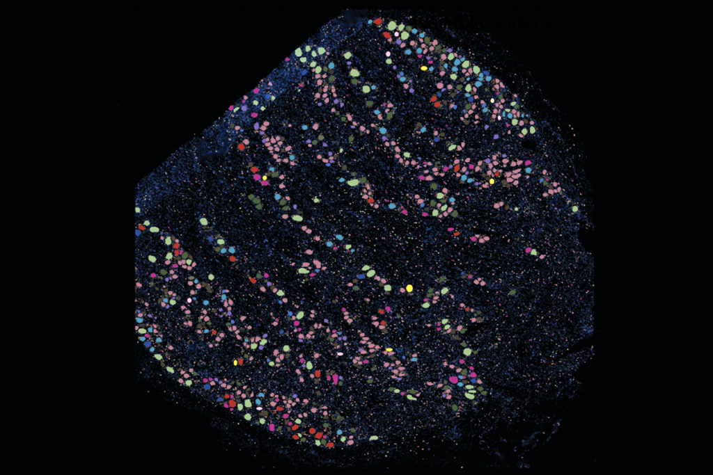
Arm atlas: A 3D map of neurotransmitter mRNA expression reveals the distribution of neurons that express dopamine (light blue), serotonin (green), the invertebrate neurotransmitter octopamine (red), and the neuropeptides FLRIamide (yellow) and bradykinin-like neuropeptide (dark blue) in the base and tip of an octopus arm.
Winters-Bostwick et al., Current Biology 2024

The arm base contains more dopamine and serotonin neurons than the arm tip, the team found. “It is interesting, because that could provide a basis for the different functions of the two arm segments,” says David Gire, associate professor of psychology at the University of Washington, who was not involved in the study. Octopuses use the arm base to crawl and push food into their mouth and use the arm tip to explore the environment, he says.

The difference could also be due to development, Winters-Bostwick says: Octopuses grow throughout their entire life, and the tissue at the tip of the arm is younger than the tissue at the base.

Crook says she would love to produce a map of the entire arm and observe how the gradient of cell types shifts across the length of it but that “it’s beyond what is possible at the moment in terms of the capacity of the microscope and the person hours involved in reconstructing it.” It took about four months to create the animated 3D reconstructions of the two tissue sections, which each account for only half a millimeter of the total length of the arm, Winters-Bostwick says. And a pygmy octopus arm can grow up to 8 centimeters long.

The team observed only one or two neurons that express gamma-aminobutyric acid (GABA), the most abundant inhibitory neurotransmitter in mammals, every few tissue slices, even though the octopus brain does contain the cell type. “It’s very different than what we see in other systems, certainly mammalian systems, where we see a lot of GABAergic neurons,” Gire says. Future electrophysiology experiments could elucidate how the octopus peripheral nervous system handles inhibition.

O

ne limitation of the work is that it “was done in one species, for technical reasons, although they did choose a good species,” Gire says. The pygmy octopus is small—the body grows to about 2 and a half centimeters—so imaging the tissue is more manageable than with larger species, Gire says. But because of its size, it does not have the same predator behaviors as larger species, which could translate to a different cellular makeup.

Gire says he would like to see similar work conducted in more species of cephalopods so researchers can do comparative studies and gain a deeper understanding of the circuitry driving different behaviors.

Some of this work already exists. The arm neural circuitry of Octopus bimaculoides shows a segmental organization oriented around the suckers, according to a preprint posted on bioRxiv in June. This pattern jibes with what Crook’s group reported in the electron microscopy dataset, says preprint author Clifton Ragsdale, professor of neurobiology at the University of Chicago. His team observed similar organization in a species of squid: The segments appear in the arms and parts of the tentacles that contain suckers, but not in the sections without.

Crook and her team are working on a range of follow-up studies: a cell-level connectome of the cluster of nerves underneath each sucker, and calcium imaging of neuronal activity in the arm.

Those experiments have been particularly exciting, Crook says. Because of the two datasets, “for the first time, when we’re looking at live imaging data, we can make assumptions about what type of cells they are, in terms of their neurochemical identity and in terms of their trajectory of their axons, which we’ve never been able to do before.”

Sign up for our weekly newsletter.

Catch up on what you may have missed from our recent coverage.