Listen to this story:
0:00
/
Person-to-person variability in brain activity might represent meaningful differences in cognitive processes, rather than random noise.
Brain imaging research may be grappling with a fresh challenge. Scanning the brain of a single person can reveal the areas they use to complete a task, although the exact pattern differs from person to person. But averaging the results across many people—as scientists often do—fails to capture some important nuances, a new functional MRI (fMRI) study suggests.
The brain tackles decision-making tasks in particular through several different categories of brain activity, rather than a single one, according to the study, published in Nature Communications in February. Across three decision-making tasks, participants’ brains differentially activated and suppressed various regions and networks in ways that could be grouped into distinct categories, or subtypes, highlighting the variability of neural signatures during behavior.
The fact that the participants performed the same tasks—yet had different activation patterns on any given one—shows “that people did different things in order to arrive at that same behavioral outcome,” says Jonas Obleser, professor of psychology at the University of Lübeck, who was not involved with the work. “Behind that what’s common, there is actually a bit more nuanced picture.”
Curiously, one of the task-related patterns involved increased activity of the default mode network (DMN), which is canonically associated with a resting or a default state. Some study participants showed DMN activity while they paid attention to the task and made decisions. These findings “complicate the picture [of the DMN], and in a very interesting way,” Obleser says. The study “sort of shoots a hole into the idea of, whenever you see the default mode network active, people are not doing the task.”
Many studies and researchers have previously hinted at the importance of individual differences in brain activity. And the person-to-person variability documented in the latest study is not unique to fMRI. For example, electroencephalography (EEG) activity patterns during a motion discrimination task could also be grouped into two categories, according to a 2023 study.
But the fMRI work shows that there is some stability to these individual differences, says Dobromir Rahnev, professor of psychology at the Georgia Institute of Technology, who led the EEG study as well as the brain activation patterns study published in February. “It’s not just meaningful variability, because people agree on that as well, but that variability may involve structured subtypes that we can actually identify and analyze.”
I
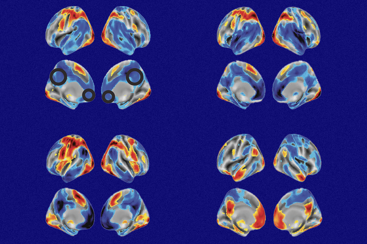
n the new study, Rahnev and colleagues applied clustering analysis to fMRI data collected from 50 adults as they completed three different decision-making tasks.For the first task—in which participants had to look at a cloud of red and blue colored dots on a screen and determine which color was more prevalent—three brain activity subtypes emerged. Subtype 1 had activation of right and left visual and parietal cortices and deactivation of the medial frontal, cingulate, temporal and right motor cortices. Subtype 2 looked similar but also had strong activation in the insula. And subtype 3 displayed activations along the DMN and deactivations in parietal and frontal areas.
When participants had to determine if the dots moved in a coherent way on a second task, their activity also coalesced into three subtypes, with similar activation patterns to those seen on the first task.
In contrast, the third task—a motion discrimination test in which participants had to judge if a proportion of white dots in a black circle were moving left or right—resulted in only two subtypes, the second of which involved increased DMN activity.
In all three tasks, one of the subtypes showed increased activity of the DMN, which is the first time DMN over-activation is linked with attention, the authors wrote in their study. The DMN’s role “might not be as simple as we thought,” says study investigator Johan Nakuci, a postdoctoral scientist in Rahnev’s lab.
Each activity subtype correlated with a different degree of behavioral performance during a task, particularly the participants’ reaction time and confidence in their decision. The different activity patterns could reflect alternate cognitive paths to perform the task, says Sarah Muldoon, professor of mathematics at the University at Buffalo, who studies brain networks. (Muldoon was Nakuci’s Ph.D. adviser but was not involved in the new work.) “When you see different spatial patterns or different regions that are activated during this task, that suggests that you’re using different thought processes, or maybe these people are thinking about it differently.”
T
he brain activity variability observed during decision-making tasks was absent during other behaviors and cognitive tasks. The fMRI signals during a working memory task, for example, looked fairly similar across all participants. This difference could be because, unlike decision-making, there are few alternative ways to approach the same problem of recall, which would result in more homogenous brain activity, Obleser speculates.Rahnev says he is not advocating for scientists to stop averaging neural signals, which he says reveals important information about general patterns. But, he says, “you are losing a lot of information when you average.”
Instead, the findings suggest that neuroscientists should move beyond merely looking at the averages of activation patterns and dig into the individual data to identify unique characteristics across subtype groups, Nakuci says. This means that neuroscientists should embrace variability, “instead of thinking of variability purely as noise.”
Next, the team wants to understand why these subtypes emerge, Rahnev says. “Where does this variability come from?” he wonders. “I don’t think that question has a single answer.”