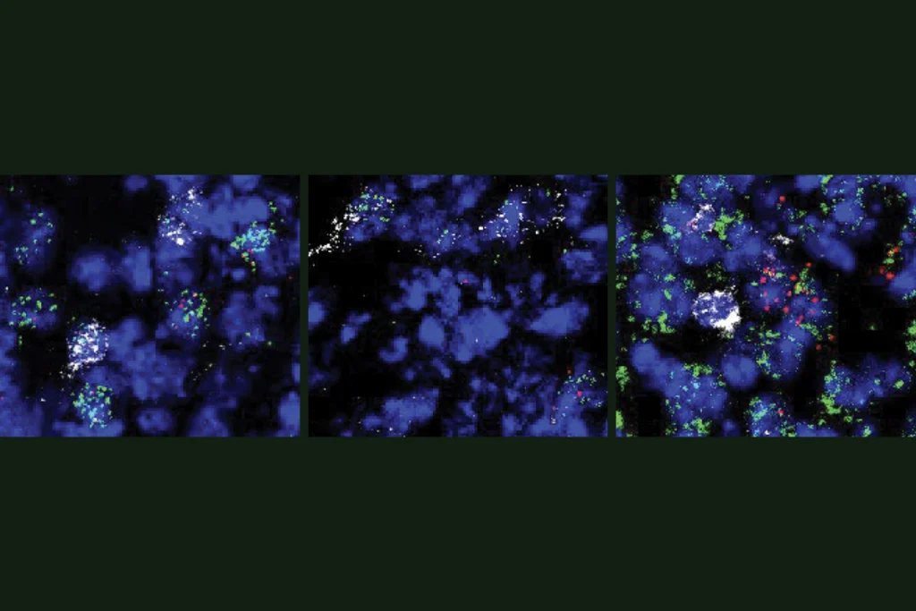
According to a half-century-old model, the striatum sends commands to motor neurons through two channels: a “go” pathway that initiates actions and a “no-go” pathway that inhibits them. But the recent characterization of two new circuits suggests other signals come into play, too.
Unlike the go and no-go pathways, which arise from an area of the striatum called the matrix, the new circuits involve striosomes, striatal structures that are neurochemically distinct from and have little communication with the matrix. These striosomal circuits, according to a new study, connect to and have opposite effects on dopamine-releasing cells in the substantia nigra, a region that neighbors the striatum—part of the basal ganglia—and may regulate movement by affecting mood and motivation.
Through their effects on dopamine levels, the striosomal circuits could modulate signals traversing the classic go and no-go pathways, which contain D1 and D2 receptors, respectively—and “may be very critical for learning,” says lead researcher Ann Graybiel, professor of brain and cognitive sciences at the Massachusetts Institute of Technology, who discovered striosomes in 1970. Because striosomes receive input from a broader range of areas, the new results point to routes by which cognitive and emotional inputs may influence movement, she says.
The field has historically lacked tools to study striosomal neurons, so the basal ganglia’s reputation as a controller of movement was based on incomplete information, says William Stauffer, assistant professor of neurobiology at the University of Pittsburgh, who was not involved in the new study. “The basal ganglia, for the last 50 years, has been a movement structure, but we know it’s much richer than that,” he says. The newly identified pathway, he adds, “should change our conception of the basal ganglia.”
“If you wait a few years, you’re going to see it in textbooks,” says Richard Courtemanche, professor of health, kinesiology and applied physiology at Concordia University, who was not involved in the work.
J
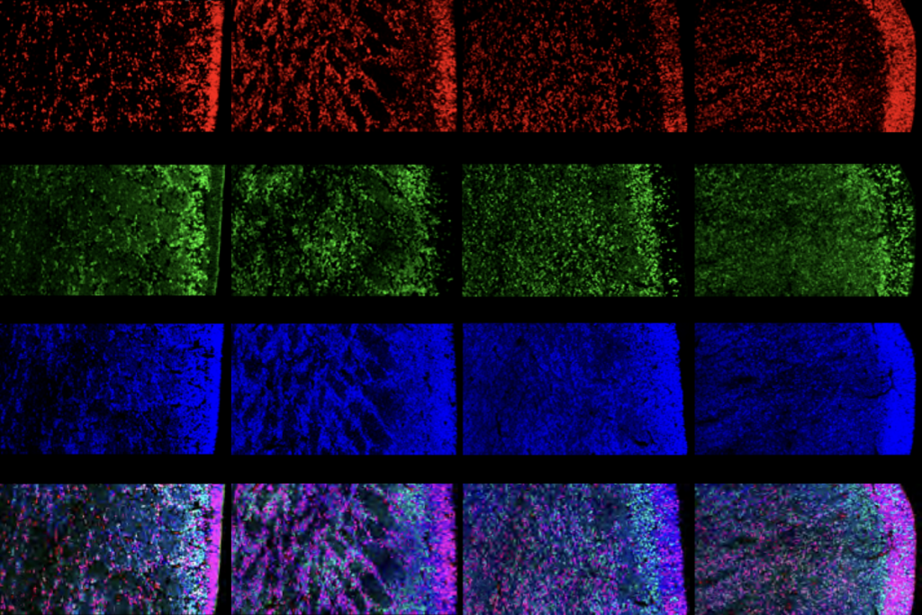
ust like the classic pathways, the striosomal circuits—one excitatory and one inhibitory—also express D1 or D2 receptors, Graybiel and her colleagues reported in 2016. The new results took 11 years of engineering to produce two mouse lines that express fluorophores to selectively mark neurons in striosomes with either D1 or D2 receptors.Cells with D1 receptors extend directly to the substantia nigra, whereas those with D2 first project onto neurons in an area called the external globus pallidus and then to the substantia nigra. The team discovered these routes by injecting the mice with a virus that travels transneuronally.
The indirect striosomal pathway is “something that just has never really been in our circuit diagram,” says Talia Lerner, associate professor of neuroscience, psychiatry and behavioral sciences at Northwestern University, who was not involved with the study. “It gives you all these ideas about how we can redraw our circuit map.”
Neurons in the two striosomal pathways behaved differently during a task in which the mice turned either left or right in a T-maze to obtain a reward. And the two striosomal circuits also differ in function, optogenetics work showed: Turning on the D1 neurons dampens dopamine release and movement, whereas turning on the D2 ones increases both outcomes. The findings were published in Current Biology in November.
The dopamine effects are the opposite of what happens in the canonical basal ganglia pathway—in which the D1 ‘go’ pathway is excitatory, and the D2 ‘no-go’ route is inhibitory—but the mechanism is “still a brake-accelerator model,” with the neurons in the striosomal pathways “actually providing a behavioral input,” Courtemanche says.
The fluorescent markers used in the study failed to flag some striosomal neurons—raising questions about whether the connections characterized in the new work are representative of the entire circuit, Lerner says. Future research should focus on creating tools to study those remaining circuits, she adds.
It will also be important to clarify the function of these striosomal pathways, and how they fit with circuits already known to control movement, Lerner says. Because striosomal pathways do not project to motor nuclei, it is likely that they influence movement through effects on motivation, she says.
Striosomes, but not the rest of the striatum, receive inputs from higher-order areas such as the cortex, so the circuits described in the new work could serve to adjust or influence certain behaviors, Graybiel says. The next steps are to identify which higher-order areas project onto the striosomes, in an effort to illuminate the circuit from cortex to striosomes to behavior, she says. “That’s a tall order.”
