Sometimes in neuroscience, as in fashion, what’s old becomes new again. Consider the glia limitans superficialis (GLS), the thin layer of cells that covers the brain and spinal cord just beneath the protective meninges. More than 100 years ago, Swedish anatomist Magnus Gustaf Retzius discovered that astrocytes form this seemingly impermeable layer, aligning their cell bodies along the brain’s surface and shooting their bushy processes into the cortex below—a pattern seen in drawings of Golgi-stained tissue from cats, dogs, rabbits and humans.
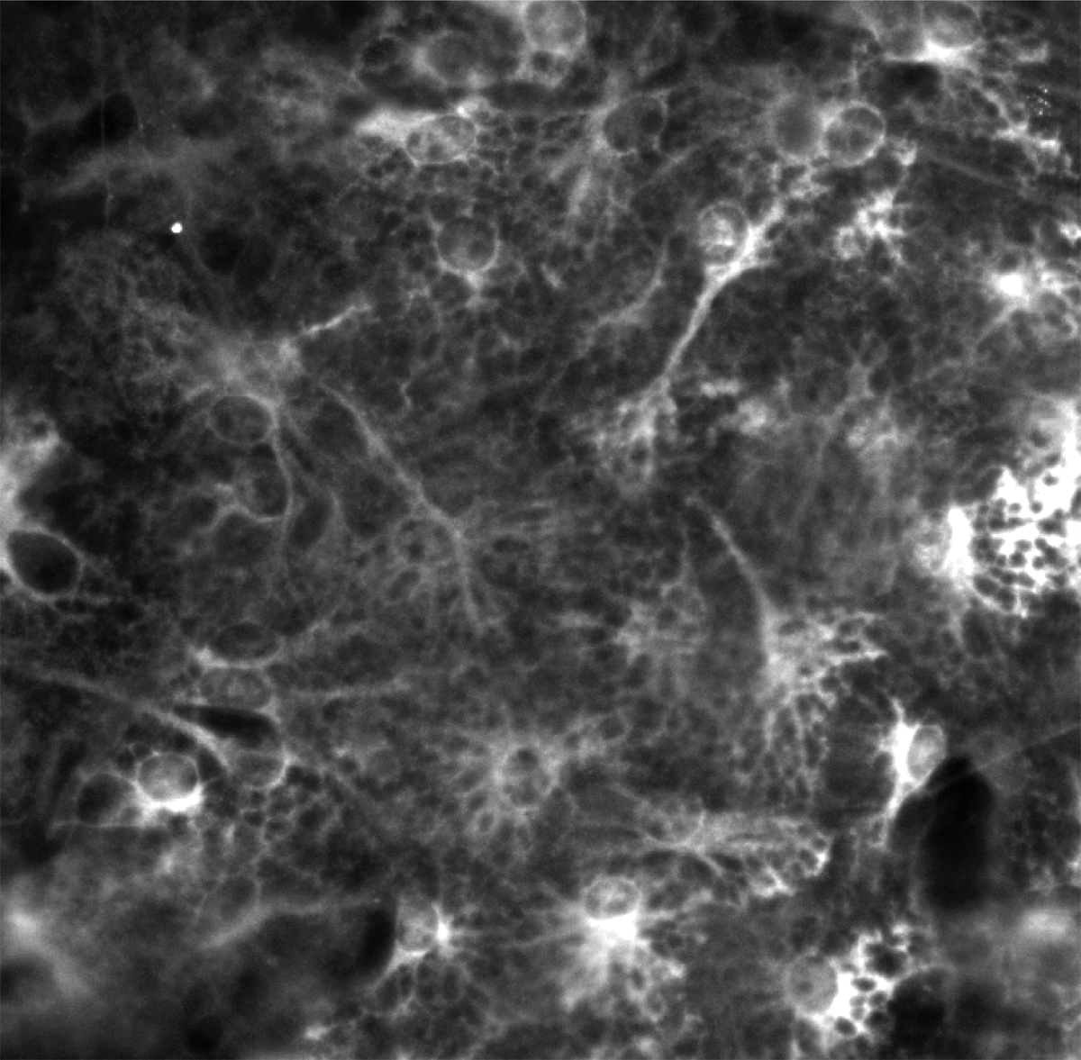
Marking territory: Astrocytes of the glia limitans superficialis differ from other types of astrocytes in that they express a gene called MYOC, as seen in this image generated through RNA in-situ hybridization.
Courtesy of Hasel et al.
Unexpected astrocyte gene flips image of brain’s ‘stalwart sentinels’
The genetic marker upends the accepted orientation of non-star-like astrocytes in the glia limitans superficialis.
By
Lauren Schenkman
28 March 2025 | 5 min read
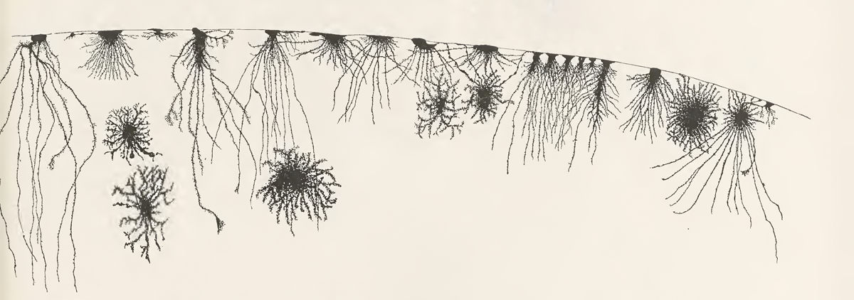
Bushy borderline: Glia limitans superficialis astrocytes appear in Retzius’s drawings of Golgi-stained brain tissue from a cat.
But in the 20th century, this picture flipped upside down: Diagrams in textbooks as recent as 2023, for example, depict GLS astrocytes with their cell bodies instead rooted in the cortex and their processes propped against the meninges, forming the sheath. In place of pathology slides, these results identified astrocytes based on their expression of astrocyte-specific genes, such as GFAP, which flags astrocyte bodies in the cortex.
New evidence from 3D light-sheet microscopy turns the picture right side up again. It reveals “a sea of cell bodies that cover the brain surface, absolutely beautiful,” in a variety of species, says Philip Hasel, group leader at the UK Dementia Research Institute in Edinburgh, who led the work, which was published in February in Cell Reports. The results, based on a reanalysis of existing data from zebrafish, mice, macaques and humans, jibe with evidence from similar reports from the past decade that “no one has listened to,” Hasel says. The new study reveals that GLS astrocytes have a unique genetic profile and are located only on the brain’s surface.
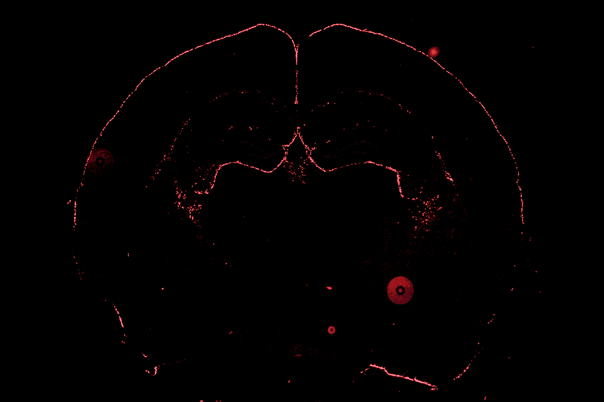
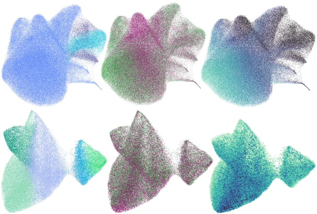
Floating army: A layer of astrocytes floats on the surface of the mouse brain, 3D light-sheet microscopy shows.
Courtesy of Hasel et al.
These “stalwart sentinels” lack the typical star shape of other astrocytes, says study investigator Shane Liddelow, associate professor of neuroscience, physiology and ophthalmology at New York University. Instead, they resemble umbrellas, with a single process shooting down into the brain like a handle.
Heads up: Astrocytes that comprise the glia limitans superficialis position their cell bodies on the surface of the brain and shoot their processes down into the interior, not the other way around, 3D light-sheet microscopy of the mouse brain reveals.
Courtesy of Hasel et al.
The results demonstrate that researchers should look beyond GFAP when studying astrocyte gene expression, Hasel says. GLS astrocytes in mice instead express high levels of the gene myocilin (MYOC), the new study shows, so staining for GFAP won’t catch them. Hasel says his team doesn’t yet know what the gene does.
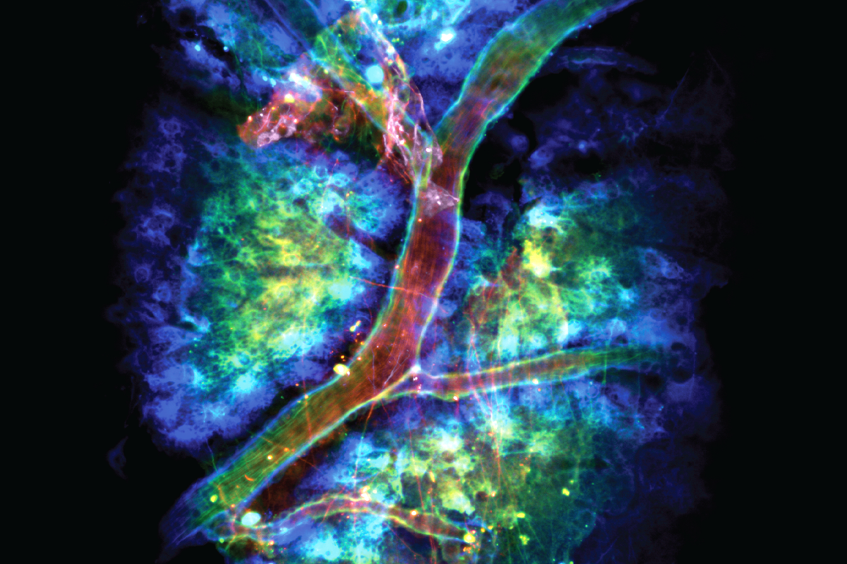
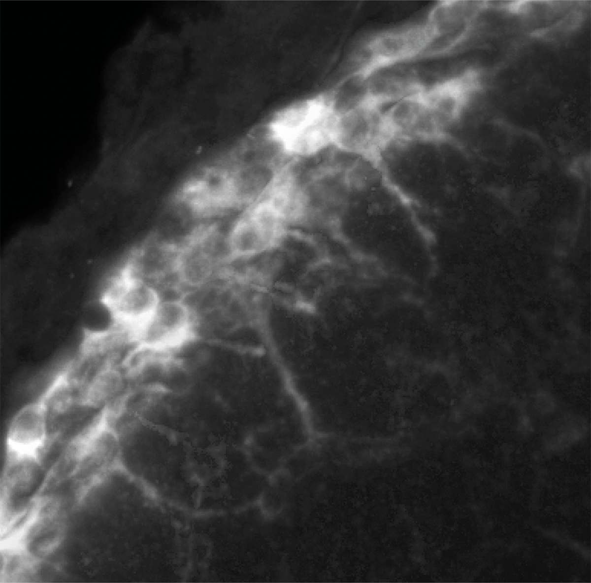
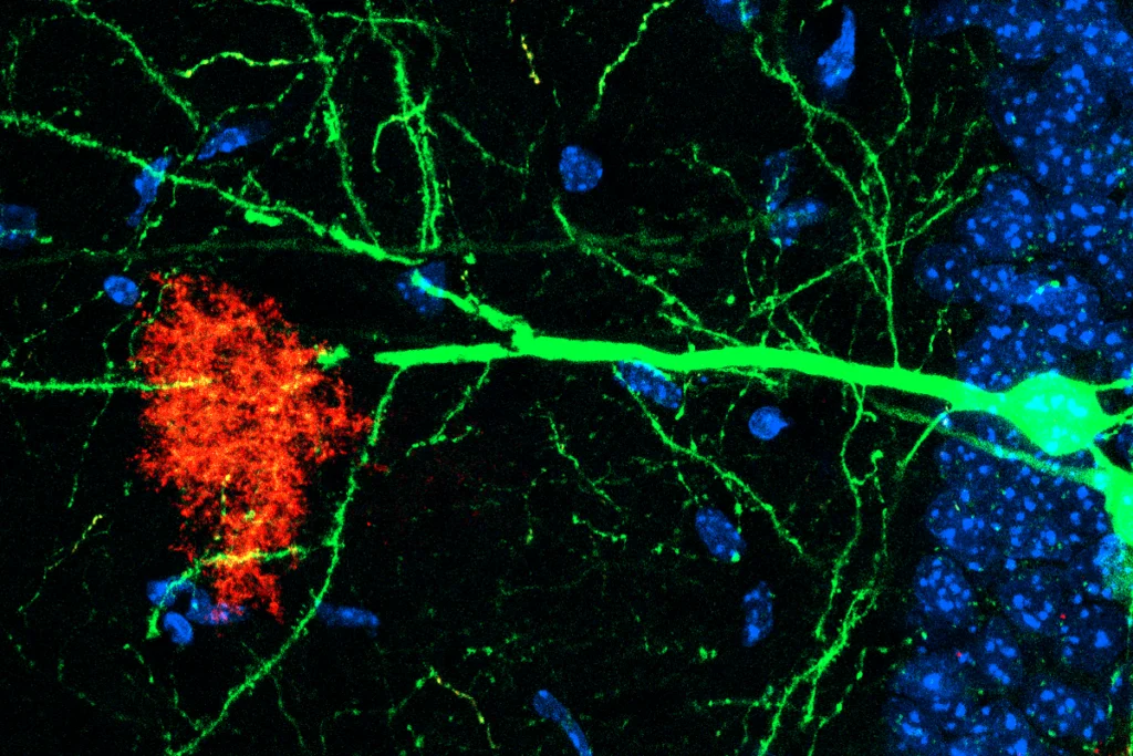
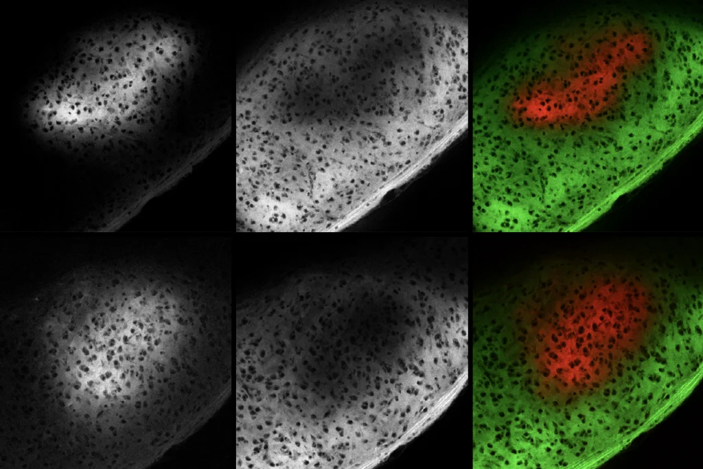
Long tail: Processes from GLS astrocytes (red) extend hundreds of microns into the cortex and crisscross over a superficial blood vessel in this image of a mouse brain.
Courtesy of Hasel et al.
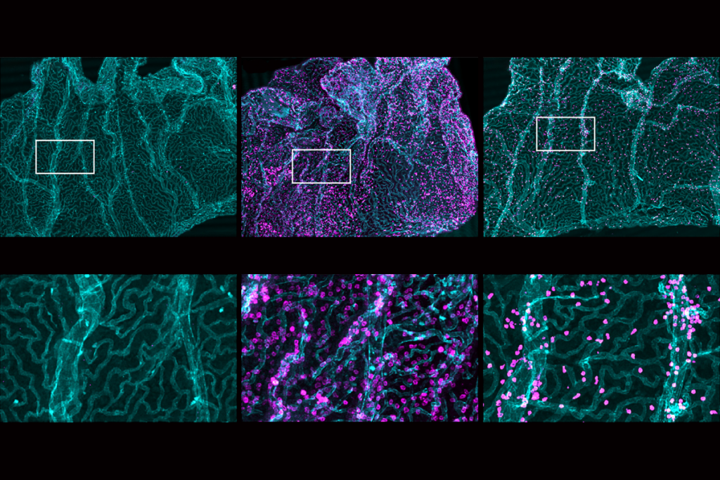
Farther down the evolutionary ladder, astrocytes with high expression of MYOC line the spinal cord of zebrafish and show the same morphology, with the cell body at the surface and processes reaching into the spinal cord.
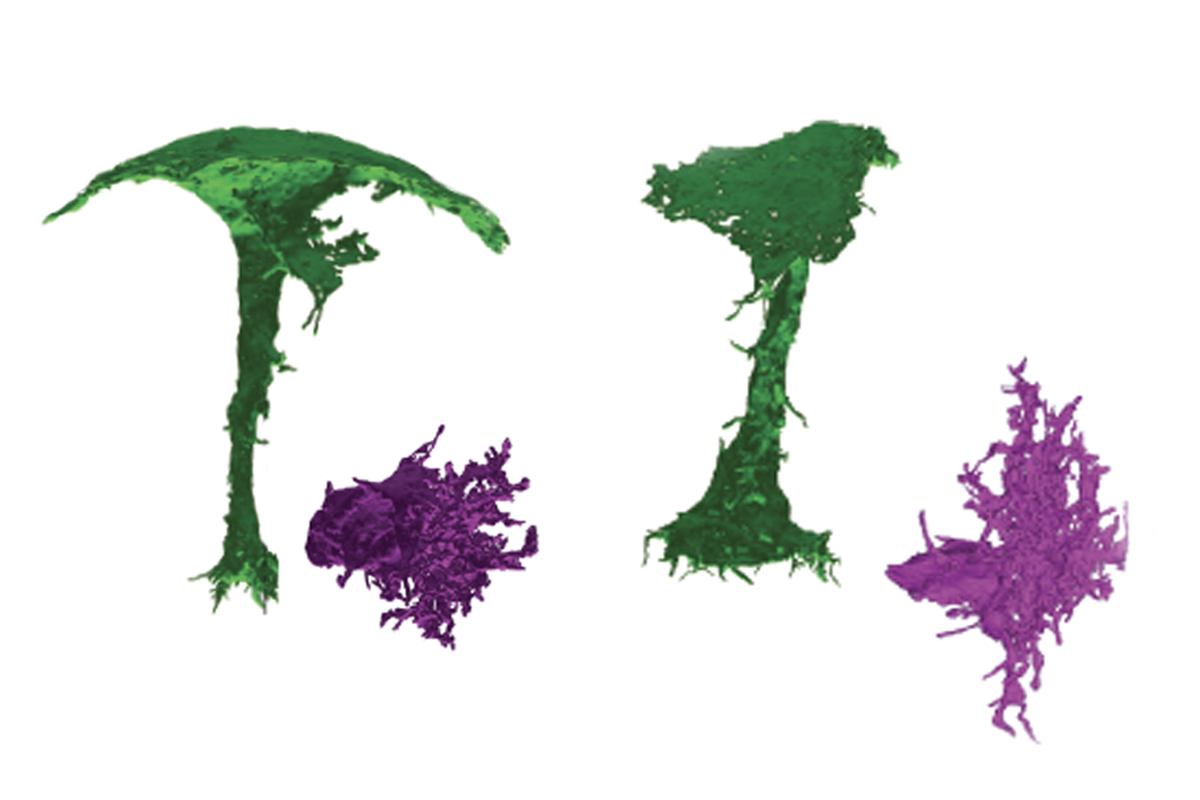
Under my umbrella: In zebrafish, astrocytes wrapping around the spinal cord (green) have the same umbrella-like morphology as rodent GLS astrocytes. By contrast, astrocytes within the spinal cord (magenta) have a more traditional star-shaped structure.
Courtesy of Hasel et al.
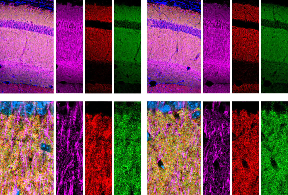
And moving up the evolutionary ladder, MYOC-rich astrocytes also coat the surface of the macaque brain and the human brain.
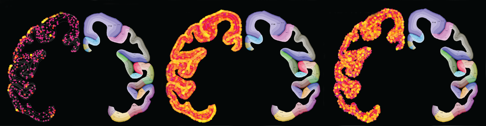
Skimming the surface: In the macaque brain, MYOC-expressing astrocytes sit on the surface (left image), whereas other astrocyte genes, ALDH1L1 and GFAP, are expressed throughout the cortex (middle and right, respectively).
Courtesy of Hasel et al.
In the mouse brain, the processes of these astrocytes extend hundreds of microns down into the cortex, but the team doesn’t yet know how this affects their function. The processes might send danger signals from the surface deep into the brain, or sniff out any intruders that have made it through, Liddelow says.
And the astrocytes are embedded in a thick layer of laminin, the team found, which could act as a protective mesh. This padding might explain why, in previous studies, circulating immune cells were found to penetrate the brain’s meninges and then apparently bounce off the brain itself. GLS astrocytes also display a gene-expression profile similar to that of an internal astrocyte reacting to a pathogen, suggesting they are already primed for action.
The new study, which entirely leveraged preexisting datasets, shows that “combining our efforts across the field can be really critical” for making new discoveries, says Sarah Ackerman, assistant professor of pathology and immunology at the Washington University in St. Louis, who was not involved in the work.
The study also demonstrates that astrocytes are even more heterogeneous than the field previously suspected, Ackerman says. It’s a lesson “to not discount something as not being an astrocyte just because of what it looks like.”
Recommended reading
Astrocytes sense neuromodulators to orchestrate neuronal activity and shape behavior
By
Claudia López Lloreda
27 June 2025 | 9 min listen
‘SNAP’ dance of astrocytes and neurons falls out of step with age, disease
By
Laura Dattaro
6 March 2024 | 6 min read
Dispute erupts over role of sticky proteins in astrocytes
By
Lauren Schenkman
10 July 2023 | 9 min listen
Explore more from The Transmitter
Immune cell interlopers breach—and repair—brain barrier in mice
By
Claudia López Lloreda
20 November 2024 | 6 min listen
Astrocytes star in memory storage, recall
By
Angie Voyles Askham
6 November 2024 | 5 min read
Microglia’s pruning function called into question
By
RJ Mackenzie
24 October 2024 | 9 min read
Cite this article: