Age-related brain changes in mice strike hypothalamus ‘hot spot’
Neuronal and non-neuronal cells throughout the brain also express genes—particularly those related to neuronal structure and immune function—differently in aged mice, according to a new atlas.
Old age is the best predictor of Alzheimer’s disease, Parkinson’s disease and many other neurodegenerative conditions. And yet, as deeply studied as those conditions are, the process of healthy brain aging is not well understood.
Without that knowledge, “how can we possibly fix something that goes wrong because of it?” asks Courtney Glavis-Bloom, senior staff scientist at the Salk Institute for Biological Sciences. “We don’t have the basics. It’s like running before we walk.”
That said, mounting evidence suggests that aging takes a particular toll on non-neuronal and white-matter cells in mice. For example, white-matter cells display more differentially expressed genes in aged mice than in younger ones, according to a 2023 single-cell analysis of the frontal cortex and striatum. And glia present in white matter show accelerated aging when compared with cells in the cortex across 15 different brain regions, another 2023 mouse study revealed.
“Different brain regions show totally different trajectories regarding aging,” says Andreas Keller, head of the Department of Clinical Bioinformatics at the Helmholtz Institute for Pharmaceutical Research Saarland, who worked on the latter study.
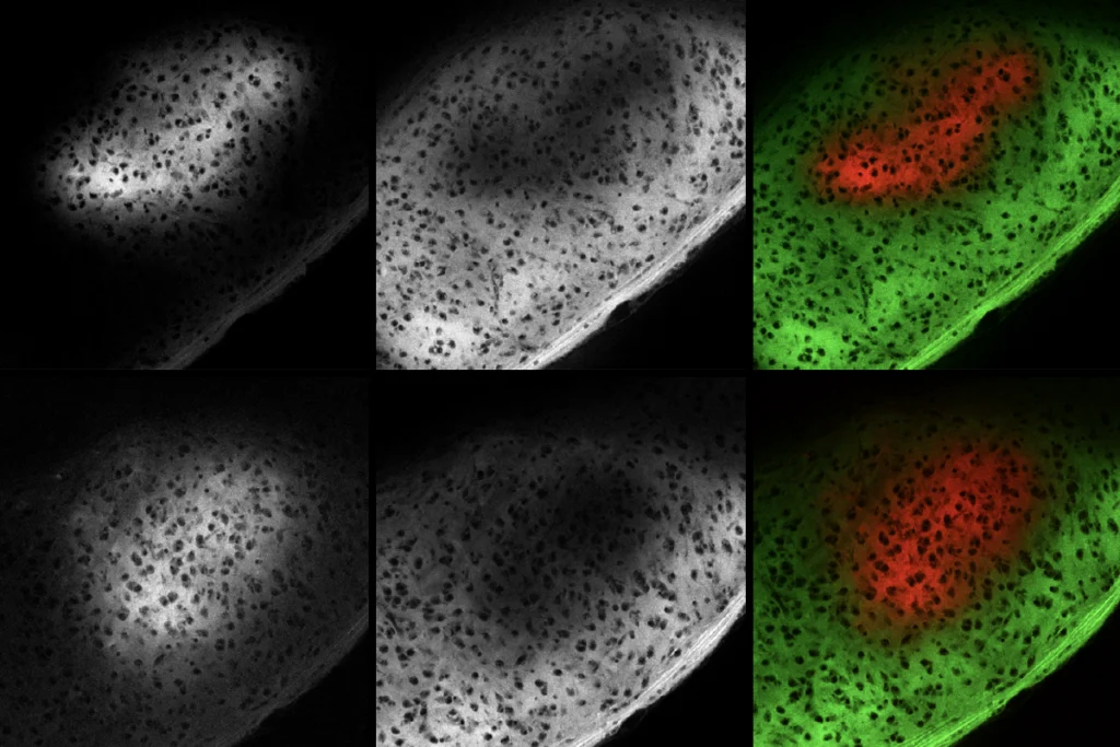
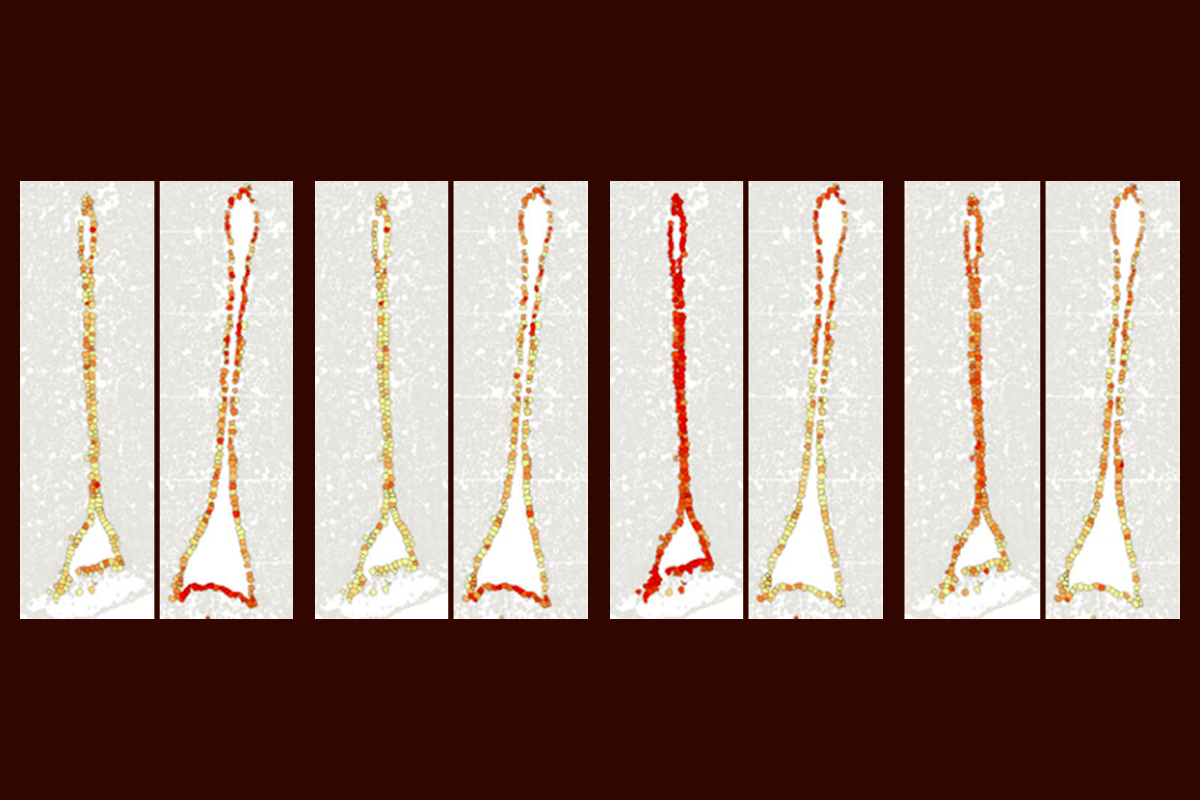
Some of the cell types with the most extensive aging-related changes in gene expression occur in a small region of the hypothalamus, according to a new single-cell mouse atlas, the largest and broadest to date. Rare neuronal and non-neuronal cell populations within this “hot spot” are particularly vulnerable to the aging process, says Hongkui Zeng, executive vice president and director of the Allen Institute for Brain Science, who led the work.
“This demonstrates the power of using the cell-type-specific approach that will identify highly susceptible, rare populations of interest in the brain,” she says.
Z
eng and her colleagues used single-cell sequencing to analyze about 1.2 million cell transcriptomes across 16 brain regions linked to aging or aging-related conditions in 2-month-old young adult mice and 18-month-old aged mice.Compared with young mice, the older mice showed a decrease in the expression of genes related to neuronal function and structure—including those linked to axon projection and cell migration—across neurons, astrocytes and oligodendrocytes. Clusters of microglia and macrophages that monitor immune activity at the brain’s barriers also showed increased inflammatory responses with aging, the team reported last week in Nature.
That result supports previous findings, Keller says. “It’s not only neurons that age, but it’s all these other cell types that are around them that maybe are even more vulnerable.”
The neuronal populations affected in the hypothalamus “hot spot,” a small region called the third ventricle, regulate metabolism and homeostasis and respond to hormones such as leptin and GLP-1. Similarly, the age-affected non-neuronal cells in the area, including ependymal cells and tanycytes, help the brain sense nutrients and hormones from the periphery. That pattern fits with the idea that aging reflects a breakdown in homeostasis, Zeng says. “We think that it’s a combination of deterioration of neuronal function, especially hormone regulation, and the increased immune response and inflammatory response that’s happening in the aging brain.”
Aging is known to alter energy homeostasis in the periphery, but the new work “puts that part of the hypothalamus as a focal point that seems to change quite dramatically with age,” says Ashley Webb, associate professor at the Buck Institute for Research on Aging, who was not involved in the study. The third ventricle, she adds, “might be an area where we need to focus—and better understand mechanistically.”
O
ne next step is to figure out whether the hypothalamus is particularly susceptible to an aging process that begins elsewhere, or whether it initiates that process. It is possible, for example, that the changes are not necessarily signs of deterioration, Webb says. “Some of these changes may actually be adaptive or compensatory, and that could actually be a good thing.”An analysis of how gene expression differs across aging male and female mice—using data that Zeng and her team collected for the atlas but did not analyze in the current paper—would also be valuable to the field, Webb says. And future atlases should expand the age range studied to include even older mice, Webb adds. “The 18-month-old time point is a little young,” roughly the equivalent of a 60-year-old human, she says.
The team plans to replicate the work in 24-month-old mice, Zeng says. “Then we’ll see if the aging is spreading into more regions, and more cell types.” She and her colleagues also plan to investigate whether epigenetic changes occur with age, she adds.
By one day targeting specific cell types that show early signs of aging, Zeng and her colleagues aim to alter that vulnerability, she says. “We want to study those cell types by manipulating them in some way—and to see if the aging process can be exacerbated or even reversed.”
But even if these signs of brain aging can be reversed in mice, whether the results will hold up in people remains unclear, Glavis-Bloom says.
“I think it’s going to be amazing to be able to eventually apply this method to the nonhuman primate brain, because mice and humans have a lot of evolutionary history between them,” she says. “What we find out about the brain in nonhuman primates is going to be much more predictive of what we should be modifying.”
Recommended reading
Aging as adaptation: Learning the brain’s recipe for resilience
Astrocytes stabilize circuits in adult mouse brain

Profiles of neurodevelopmental conditions; and more
Explore more from The Transmitter
Early trajectory of Alzheimer’s tracked in single-cell brain atlases
